
Page 31 - Journal of Structural Heart Disease Volume 3, Issue 1
P. 31
Original Scienti c Article
24
Video 24. “Cobra”-shape dis guration of the left disc. View sup- plemental video at http://dx.doi.org/10.12945/j.jshd.2016.005.16. vid.24.
Video 25. A oating device in the left atrium following its re- lease. View supplemental video at http://dx.doi.org/10.12945/j. jshd.2016.005.16.vid.25.
Video 26. A oating device in the left atrium following its re- lease. View supplemental video at http://dx.doi.org/10.12945/j. jshd.2016.005.16.vid.26.
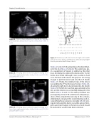
Figure 6. Simultaneous left ventricle (LV) and right ventricle (RV) pressure tracings during catheterization demonstrating higher diastolic pressure in the RV than in the LV.
(Videos 23 and 24). Following release, the device oat- ed in the LA (Videos 25 and 26). The patient was giv- en a supplement of heparin in addition to the initial dose. An attempt to retrieve the device with a 15-mm Andra snare failed. Although it was possible to hold on to the hub of the device, the grip was not strong enough to pull the device back into the 12F sheath (Video 27). Finally, the retention hub of the device was grabbed by 7F Cordis biopsy forceps (Video 28), allowing the right disc to be retrieved into the sheath (Video 29). The left disc was then approximated to the IAS, and the device was successfully deployed in the defect (Videos 30 and 31). The entire procedure was prolonged by 25 min. TTE con rmed an adequate po- sition of the occluder the next day. In this scenario, attempting to recapture the connecting hub and ac- complishing the procedure is desirable [29, 30]. Snar- ing and removing the device is another option. Emer- gent open-heart surgery is the last resort and should be reserved for unsuccessful device retrieval.
Journal of Structural Heart Disease, February 2017
Volume 3, Issue 1:15-27
