
Page 36 - Journal of Structural Heart Disease Volume 3, Issue 1
P. 36
29
Case Report
a calci ed substrate also increases the risk of malposi- tioning. Furthermore, there is a risk of residual aortic regurgitation, resulting in a high rate (18.8%) of need for second valve implantations [1].
Nevertheless, several reports have described the use of TAVR in patients with NAVR. In a registry study of 43 patients from 14 di erent countries, Roy et al. demonstrated the feasibility of CoreValve implan- tation in extreme operative risk patients with NAVR without aortic stenosis [1]. The implantation of a Cor- eValve was successful in 97.7% of cases; however, the Valve Academic Research Consortium-de ned proce- dural success rate was only 74.4% due to the need for a second valve in some cases. The Italian CoreValve multicenter registry con rmed its feasibility in 26 pa- tients and showed that patients with NAVR are usu- ally younger than those undergoing TAVR for aortic stenosis [2]. In this report, we describe a challenging case of implanting a CoreValve for NAVR and a repro- ducible solution.
Case Presentation
The patient was a 38-year-old male with pulmonary valve dysplasia associated with Noonan’s syndrome and hypertrophic cardiomyopathy. The patient un- derwent pulmonary valvotomy and myomectomy at 17 months of age followed by a pulmonary homo- graft for severe pulmonary insu ciency at 20 years of age. With a history of multiple episodes of infective endocarditis on the aortic valve, he subsequently de- veloped severe aortic insu ciency and was referred to us for possible TAVR. Due to signi cant frailty, poor pulmonary function, and prior sternotomies, he was considered a candidate for TAVR. His STS score for mortality or morbidity was calculated as 24.83%.
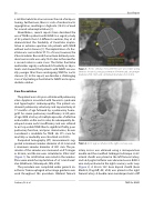
Computed tomography (CT) measurements sug- gested a minimum annulus diameter of 23.3 mm and a maximum annulus diameter of 30.1 mm. The pe- rimeter of the annulus was measured on CT images as 88 mm, and the area was calculated as 596.2 mm2 (Figure 1). No calci cation was noted in the annulus. Plans were made for implantation of a 31-mm CoreV- alve (Medtronic, Minneapolis, MN, USA).
The procedure was completed under general an- esthesia. Transesophageal echo image guidance was used throughout the procedure. Bilateral femoral
Figure 1. Aortic annulus measurements pre procedure using 3mensio showed a annulus perimeter of 88 mm and a maximum diameter of 30.1mm and a minimum diameter of 23.3 mm.
Figure 2. A 6-F pigtail catheter in the right coronary cusp.
artery access was obtained using a micropuncture needle and the modi ed Seldinger technique. A 6-F arterial sheath was placed in the left femoral artery, and a 6-F pigtail catheter was advanced over a 0.035 J wire and positioned in the right coronary aortic cusp (Figure 2). A 28-cm 18-F Gore dryseal sheath (Gore Medical, Flagsta , AZ, USA) was placed in the right femoral artery. A double curve Lunderquist extra sti
Sunkara, N. et al.
TAVR for NAVR
