
Page 18 - Journal of Structural Heart Disease Volume 3, Issue 2
P. 18
Case Report
44
visited our outpatient clinic. He was diagnosed at 43 years of age with secundum ASD requiring surgical repair but had rejected treatment at that time. At 76 years of age, he was referred from a community hos- pital to our institute due to shortness of breath on exertion. By this time, surgical intervention was no longer indicated because echocardiography revealed severe pulmonary hypertension (estimated right ven- tricular systolic pressure, 102 mmHg). He was treated conservatively with heart failure medications such as furosemide, digoxin, and carvedilol. However, be- cause his symptoms had worsened considerably over the past several years, he was referred to our institute again and hospitalized while alternative treatments were considered.
Although his vital signs were stable (blood pres- sure, 125/69 mmHg; pulse rate, 69 beats per minute with irregularity), his transcutaneous oxygen satura- tion (SatO2) in room air had decreased to 95%. During a 6-min walk test, his SatO2 on 1 l/min of nasal oxygen declined to 88%. On physical examination, his jugu- lar veins were dilated, and facial and pretibial edema were observed. Rales were audible in both lower lung elds. His heart sounds revealed xed splitting of the second heart sound and a systolic murmur at the sec- ond intercostal space and left-sternal border. Labora- tory data showed a total bilirubin of 1.3 mg/dl and B-type natriuretic peptide of 407 pg/ml. A chest X-ray revealed cardiomegaly, enlargement of the bilateral pulmonary artery, and pleural e usion (Figure 1). An electrocardiogram showed atrial brillation and com- plete right bundle branch block. Transthoracic echo- cardiography (TTE) revealed enlargement of the right heart, hypoplasty of the left ventricle (Figure 2A), and
massive pericardial e usion (Figure 2B). In addition, there was an ostium secundum-type ASD with left- to-right shunting and moderate tricuspid regurgi- tation on transesophageal echocardiography (TEE) (Figure 2C). The size of the defect and the rims were also measured by TEE. His ASD showed a maximal size of 36 × 37 mm, and each rim was long enough for device closure (Figure 3). Cardiac catheterization revealed Qp/Qs of 4.6, pulmonary artery pressure of 93/35/52 mmHg, and pulmonary vascular resistance
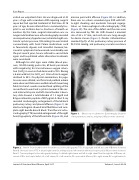
Figure 1.
Chest x-ray on admission. Cardiomegaly, dilatation of the pulmonary arteries, and pleural e usion were observed.
Figure 2. Panel A. Four-chamber view of transthoracic echocardiography (TTE). A large right and left atria and ASD were observed. Panel B. Short-axis view of TTE. A dilated right ventricle and hypoplastic left ventricle with massive pericardial e usion were observed. Panel C. Transesophageal echocardiography ndings. A dilated right atrium and left-to-right shunt ow through the ASD were ob- served. ASD = atrial septal defect; LA = left atrium; LV = left ventricle; PE = pericardial e usion; RA = right atrium; RV = right ventricle.
Journal of Structural Heart Disease, April 2017 Volume 3, Issue 2:43-48
