
Page 19 - Journal of Structural Heart Disease Volume 3, Issue 2
P. 19
45
Case Report
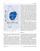
Figure 3. Transthoracic echocardiography ndings of atrial sep- tal defect (ASD). The ASD occupies large part of the atrial septum. SVC = Superior vena cava; IVC = Inferior vena cava.
temporarily, we observed that pulmonary capillary wedge pressure and left ventricular end diastolic pressure did not rise markedly. Finally, we successful- ly implanted a 38-mm ASO, which was the maximum size available in Japan at the time (Figure 5). No pul- monary edema occurred after ASO implantation, and the patient’s symptoms improved without oxygen administration. When cardiac catheterization was performed on postoperative day 13, there was no evidence of residual shunting, and pulmonary artery pressure had decreased to 63/20/33 mmHg. As peri- cardial e usion was still present after successful ASD closure, pericardiocentesis was performed, and 1,500 ml of uid was removed. A PDE5 inhibitor (sildena l 60 mg), endothelin receptor blocker (ambrisentan 5 mg), and PGI2 analog (beraprost 120 μg) were admin- istered to treat the remaining pulmonary hyperten- sion. The patient was subsequently discharged from the hospital on postoperative day 41 and returned home on foot.
Three months after ASO implantation, the patient was re-hospitalized due to dyspnea on exertion be- cause pericardial e usion appeared again, even af- ter pericardiocentesis and intensive medical therapy (Figure 6). His pulmonary artery pressure was still high at 47/26/35 mmHg. Therefore, surgical pericar- diostomy was performed 6 months after ASD closure. Finally, his left ventricular shape became round and enlarged (Figure 7), and pulmonary artery pressure decreased to 34/16/24 (Figure 6). Finally, he was dis- charged again and became asymptomatic.
Discussion
We successfully treated a patient with a giant ASD along with extreme pulmonary hypertension and massive pericardial e usion percutaneously with an ASO. Therefore, this case shows that appropriate and timely pre- and postoperative medical therapy com- bined with ASD closure can be e ective.
In patients with signi cant ASD, left ventricular pre- load is reduced and a reduction of LV volume is ob- served over time [4]. In these cases, closure of an ASD with a large left-to-right shunt can increase left ventric- ular in ow, leading to left ventricular heart failure [5]. To avoid this catastrophic condition, two diuretics and an inotropic agent were administered intravenously for
(PVR) of 8.3 Wood units. His PVR index was 5.4 Woods units · m2, and pulmonary-to-systemic blood pressure ratio was 0.87.
Mainly due to his frailty, percutaneous treat- ment with an Amplatzer septal occluder (ASO) was planned. Two di erent diuretics, a PDE3 inhibitor (milrinone 0.2 μg/kin/min) and nasal oxygen, were administered as preoperative therapy in preparation for acute decompensation (Figure 4). After 1 month of medical treatment, we performed percutaneous device closure for atrial septal occlusion. We per- formed the operation while monitoring intra-cardiac pressure. When we occluded the defect with an ASO
Kikuchi, N. et al.
Percutaneous Device Closure for Giant ASD and PE
