
Page 31 - Journal of Structural Heart Disease Volume 3, Issue 2
P. 31
57
Case Report
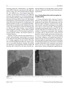
Following diagnostic catheterization, an Amplatzer super-sti (St. Jude Inc.) was placed in the right pul- monary artery, and a PTS-X 30-mm balloon (Numed Inc.) was in ated across the tricuspid bioprosthe- sis with a 21-mm waist noted (Figure 2A). A Melody valve was then mounted on a 22-mm Ensemble bal- loon-in-balloon delivery system and deployed within the Hancock II bioprosthesis (Figure 2B). Subsequent hemodynamic assessment showed a reduction in the mean in ow diastolic gradient to 2 mmHg (Figure 1B) without evidence of intra- or paravalvular leak (PVL). The patient was extubated in the catheterization lab- oratory and discharged to home the following day on daily 325 mg aspirin therapy. A post-operative echocardiogram revealed a mean in ow gradient of 3.5 mmHg and trace TR. The patient reported in- creased physical stamina and denied dyspnea on moderate exertion.
However, 8 months after the valve implantation, a routine echocardiogram demonstrated an increase in the diastolic transvalvular mean Doppler gradient to 11 mmHg, suggestive of valve stenosis. The valve was not well visualized, but there was clinical suspi- cion of a possible thrombotic etiology. The patient was initiated on anticoagulation with rivaroxaban 20 mg daily with a reduction in the mean diastolic gra-
dient by Doppler to 4 mmHg within 3 weeks, and the valve has continued to function well 14 months post- implantation.
Case 2: Failing Native Valve with Incomplete An- nuloplasty Ring
A 59-year-old female with a history of atrial - brillation and rheumatic mitral valve (MV) stenosis underwent bioprosthetic MV replacement and the Maze procedure at 46 years of age. At 57 years of age, she received a redo MV replacement for prosthetic valve stenosis and TV repair with a 30-mm Edwards incomplete annuloplasty ring for severe TR. She de- veloped high-grade atrioventricular block and also received a transvenous permanent dual chamber pacemaker with an atrial lead and a ventricular lead placed in a lateral cardiac vein via the coronary sinus to avoid crossing the TV. Within 1 year, she developed heart failure symptoms and required up-titration of diuretic therapy and home intravenous dobutamine. She had progressive renal failure and severe recurrent TR requiring hospitalization and initiation of multiple inotropic medications and continuous veno-venous dialysis. She was also diagnosed with congestive he- patic cirrhosis. She was evaluated for surgical valve re-
AB
Figure 2. Panel A. Balloon in ation across the stenotic prosthesis indicated a 21-mm waist. Panel B. Post-placement of a 22-mm Melody valve.
Zhou, S. et al. Percutaneous Tricuspid Valve Replacement
