
Page 33 - Journal of Structural Heart Disease Volume 3, Issue 2
P. 33
59
Case Report
Association (NYHA) class IV prior to the procedure to NYHA class II within 3 months. However, increasing se- verity of the lateral PVL precipitated worsening heart failure symptoms and volume overload. Therefore, she underwent repeat catheterization 10 months
after valve replacement with successful occlusion of the lateral PVL under intracardiac echo guidance (Figure 5A). Follow-up echocardiography at 3 and 6 months showed stable valve function and mild lateral and medial PVL (Figure 5B).
AB
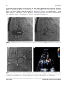
Figure 4. Panel A. Balloon in ation across the annuloplasty ring indicated a 27-mm waist. Panel B. Post-placement of a 29-mm Sapien XT valve.
AB
Figure 5. Panel A. Post-deployment of additional Amplatzer vascular plugs to treat worsening lateral paravalvular leak (PVL) in addi- tion to medial plugs from initial catheterization. Panel B. Transthoracic echocardiogram with Doppler post-deployment of additional vascular plugs showed reduced PVL. Trace central regurgitation was noted. RV = right ventricular; TV = tricuspid valve.
Zhou, S. et al. Percutaneous Tricuspid Valve Replacement
