
Page 34 - Journal of Structural Heart Disease Volume 3, Issue 3
P. 34
87
Meeting Abstracts
PERCUTANEOUS BALLOON VALVULOPLASTY FOR SEVERE PULMONARY STENOSIS IN INFANTS: A 10- YEAR INSTITUTIONAL EXPERIENCE AND LONGTERM OUTCOME
Fang Liu, Xuecun Liang, Lin Wu, Lan He, Lu Zhao, Pengjun Zhao, Guo-Ying Huang
Pediatric Heart Center, Children’s Hospital of Fudan University, Shanghai, China
Background: Although percutaneous balloon pulmonary valvu- loplasty (PBPV) is the primary treatment for signi cant pulmonary valvular stenosis, it’s widely considered to be di cult and relatively high risk for younger and severe stenosis children.
Objective: We retrospectively reviewed and analyzed the immediate and long-term outcome and safety of PBPV in neonates and infants with severe or critical valvular pulmonary stenosis.
Methods: One hundred and nine patients aged 2d~3y with critical or severe pulmonary valve stenosis admitted to our hospital from January 2005 to December 2014 underwent balloon valvuloplasty. Among them, 21 neonates had critical pulmonary stenosis, who had a tripartite right ventricle with moderate to severe tricuspid regur- gitation (TR). Severe TR was seen in 12 and moderate TR in 6 out of other 88 patients of over one month of age. Right ventricular systolic pressure in all patients was equal to or greater than systemic pressure. 53 patients had PFO or small ASD with right-to-left or bi-directional shunt, 10 patients had PDA, 1 patient had multiple small muscular ventricular septal defects, and 1 patient had atrial septal defect, who had undergone the ASD occlusion two-year later. Dilatation with 2 balloons sequentially in one procedure was performed in 12 patients and dilatation with 1 balloon in the other patients.
Results: The pulmonary valvuloplasty was successfully performed in 105 of the 109 patients, and the dilatation success rate was 96.3%. In the four failure patients, balloon catheter could not be manipulated to cross the pulmonary valve in three patients, cardiac tamponade occurred in one patient. Immediately after dilatation, the systemic pressure gradient from right ventricle to pulmonary artery decreased from 50~132 (76.25±23.7) mmHg to 4~96 (25.29±19.2) mmHg (P<0.001). No signi cant complications in all patients during or post dilation except cardiac tamponade in one. During a 12 months to 9.6 years follow-up (mean 5.01 years), data showed that: (1) pressure gradient crossing pulmonary valve measured by echocardiography further decreased or remained stable in 103 cases, except one neo- nate and three infants, whose pressure gradient gradually increased, and needed a second dilatation and good results were gained. Re-dilatation rate was 3.73% (4/107). No case needed further surgery; (2) Tricuspid regurgitation reduced in all patients except for three whose RV were dysplasia; (3) Mild pulmonary regurgitation was seen in most patients post-dilatation, except moderate in six and severe in one. (4) All 10 PDAs closed spontaneously in 3~6 months of follow-up and muscular VSDs were closed as well in 3 months of follow-up. (5) All patients were doing well and were asymptomatic and acyanosis.
Conclusions: Balloon pulmonary valvuloplasty (BPV) is safe and e ec- tive in attaining both immediate and long term reduction of pulmo- nary valvular gradients and is currently the preferred therapeutic
modality for valvular PS even in small baby patients with severe or critical stenosis.
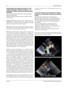
A CASE OF SEVERE AS WITH SEVERE LVOT STENOSIS SUCCESSFULLY TREATED BY TAVR AND SUBSEQUENT PTSMA
Shohei Moriyama1, Takeshi Arita1, Taku Yokoyama1, Hiromichi Sonoda3, Akira Shiose3, Yasuhiro Oga2, Yusuke Takahara2, Keiji Oi2, Ken-ichi Hiasa2, Kazumasa Fujita1, Kei Irie1, Hirotaka Noda1, Mitsuhiro Fukata1, Keita Odashiro1, Koichi Akashi1
1 Division of Hematology/Oncology/Cardioangiology, Department of Medicine, Kyushu University Hospital
2 Department of Cardiovascular medicine, Kyushu University Graduate school of medical sciences
3 Department of Cardiovascular surgery, Kyushu University Graduate School of Medicine
Aortic stenosis (AS) causes left ventricular hypertrophy which some- times leads to left ventricular out ow tract stenosis (LVOTS) like hypertrophic obstructive cardiomyopathy.
Hijazi, Z
20th Annual PICS/AICS Meeting Abstracts
