
Page 44 - Journal of Structural Heart Disease Volume 3, Issue 4
P. 44
131 Case Reports
AB
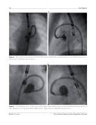
Figure 4. Agilis sheath retrogradely placed into the left ventricle and de ected toward the pulmonary valve. (Panel A) Anterior-pos- terior projection. (Panel A) Lateral projection.
AB
Figure 5. A 6-F multipurpose A-1 catheter advanced through the Agilis sheath and positioned immediately distal to the pulmonary valve apparatus for an angiogram. (Panel A) Right anterior oblique projection. (Panel B) Lateral projection.
Rhodes, J. F. et al. Transcatheter Pulmonary Value Stump Device Closure
