
Page 45 - Journal of Structural Heart Disease Volume 3, Issue 4
P. 45
Case Reports 132
AB
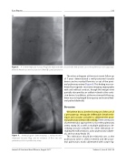
Figure 6. A 12-mm Amplatzer Vascular Plug fully deployed with a waist in the mid-portion across the pulmonary valve apparatus. (Panel A) Anterior-posterior projection. (Panel B) Lateral projection.
The echocardiogram at the most recent follow-up of 7 years demonstrated a well-positioned vascular device and no residual ow into or out of the proxi- mal pulmonary artery (Figure 8). This nding was con- rmed by magnetic resonance imaging angiography with and without contrast, though the images were partially obscured by an artifact related to the occlu- sion device. In addition, at the most recent follow-up, there was no leg length discrepancy and normal fem- oral pulses bilaterally.
Discussion
We believe this to be the rst report of the use of a percutaneous retrograde de ectable sheath tech- nique and vascular occluder to obliterate the proxi- mal pulmonary artery in this setting. There are reports of perventricular approaches to close the pulmonary artery stump [8] as well as multiple publications de- scribing vascular occluders for other indications in- cluding AV malformations, aorto-pulmonary collater- als, and coronary stula [9].
This indication should be relatively rare, as the pulmonary valve is often resected and the proxi- mal pulmonary trunk obliterated with suture lig-
Figure 7.
Ventriculogram demonstrating a well-positioned Amplatzer Vascular Plug and no evidence of ow into the pulmonary valve or pulmonary artery.
Journal of Structural Heart Disease, August 2017
Volume 3, Issue 4:128-134
