
Page 39 - Journal of Structural Heart Disease Volume 4, Issue 1
P. 39
Case Report 30
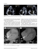
Figure 2. Echocardiogram images demonstrating right atrial volumes of 186 ml before device closure (Panel A), 160 ml the day after the procedure (Panel B), and 94 ml 8 days after the procedure (Panel C).
was discharged the next day after echocardiography showed excellent positioning of the device and no evidence of pericardial e usion.
The patient returned for routine follow-up 8 days later with complaints of palpitations and returned again 12 days later for an urgent visit with continued palpitations as well as chest pain and pressure. Re- peat chest X-ray demonstrated a profound decrease
in right atrial size (Figure 1B). Review of the patient’s transthoracic echocardiogram demonstrated a de- crease in right atrial volume from 186 ml pre-proce- dure to 160 ml the day after the procedure and 94 ml 8 days after the procedure (Figure 2).
Despite aggressive medical treatment with non-steroidal anti-in ammatory medication (i.e., opi- ates and steroids), he continued to have daily symp-
Figure 3. Computed tomography scan demonstrating the right atrial disc “digging” into the posterior wall of the right atrium and distortion of the right atrial wall (arrow). Panel A. Posterior right atrial wall distortion at the level of the pulmonary veins. Panel B. An- terior view with continued right atrial wall distortion (arrow).
Journal of Structural Heart Disease, February 2018 Volume 4, Issue 1:28-32
