
Page 40 - Journal of Structural Heart Disease Volume 4, Issue 1
P. 40
31
Case Report
toms. ECG demonstrated a low atrial rhythm with pauses greater than 2 s and brief runs of atrial ut- ter. ECG did not demonstrate pericardial e usion, but ECG-gated cardiac CTA showed the right atrial disc “digging” into the posterior wall of the right atrium (Figure 3).
Given his persistent symptoms and rhythm distur- bance, we decided that device removal and surgical closure of the ASD was the best course of action. Sub- sequent explantation of the device demonstrated distortion of the right atrial disk (Figure 4). The patient also had a pacemaker placed for sinus node dysfunc- tion and has had ablations for intraatrial re-entrant tachycardia.
Discussion
Transcatheter closure of secundum ASDs with var- ious occlusion devices has become a standard of care in most pediatric and adult patients due to greater safety and e cacy, shorter recovery duration, and de- creased hospitalization time compared with surgical closure [1]. Complications are rare with transcatheter devices, and most complications that require surgical removal and ASD patch closure are secondary to de- vice embolization [2]. Rhythm abnormalities are also reported, with supraventricular tachycardia and atrio- ventricular block being the most common [1].
Right-sided remodeling following ASD closure re- mains di cult to assess given the geometric shape of the right atrium and right ventricle. Major deforma- tional and geometrical changes of the right ventricle are completed in 24 hours, but remodeling continues for several months to years [2]. Echocardiography data using strain rate values and tricuspid annular plane systolic excursion suggest an immediate im- provement in left ventricular function and decrease in right ventricular function, likely secondary to load- ing conditions [3-6]. Changes in the size and shape of the atria are not well described.
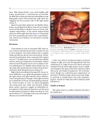
Figure 4.
Surgical exposure of the right atrium revealed a dis- torted right atrial disc. Dashed lines demonstrate the course of the device in the right atrium. The arrow corresponds to the area of atrial distortion demonstrated on the computed tomography scan in Figure 3. SVC = superior vena cava; IVC = inferior vena cava.
In this case, chest X-ray demonstrated a profound change in right atrial size. We hypothesize that the combination of a large 38-mm ASO device and rapid change in the size of the right atrium caused a signi - cant change in the shape of the device that provoked chest pain and rhythm disturbances. The conforma- tional change was con rmed upon explantation of the device. Therefore, larger devices may have a high- er potential to change shape and cause symptoms. Caution should be employed when utilizing larger ASD devices.
Con ict of Interest
The authors have no con ict of interest relevant to this publication.
Comment on this Article or Ask a Question
Reardon L. C. et al.
Rapid Right Atrial Reduction after ASD Closure
