
Page 31 - Journal of Structural Heart Disease Volume 4, Issue 2
P. 31
Case Report 52
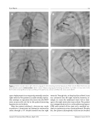
Figure 1. Panels A and B. Initial right ventricle angiogram demonstrating severely hypoplastic RVOT in both (Panel A) frontal and (Panel B) lateral planes (black arrows). Panels C and D. Repeat angiography following placement of a 4-mm coronary stent in the RVOT demonstrating (Panel C) improved lling of the pulmonary arteries and (Panel D) a more patent RVOT.
upon deployment it too migrated proximally into the right ventricle. The patient was stable initially, but fur- ther attempts to reposition the stents into the RVOT were unsuccessful and led to the patient becoming hypotensive and acidotic.
After the patient stabilized, a decision was made to perform a sternotomy and attempt to retrieve the stents through a 9-F sheath inserted into the right
ventricle. Through this, an Amplatz Goose Neck Snare was advanced into the right ventricle; however, at- tempts to snare the embolized stents led to dam- age to the right ventricular myocardium. The patient was emergently placed on cardio-pulmonary bypass (CPB) with retrieval of the stent and a modi ed sys- temic-to-pulmonary artery shunt performed. Despite this, the patient subsequently could not be weaned
Journal of Structural Heart Disease, April 2018
Volume 4, Issue 2:50-55
