
Page 32 - Journal of Structural Heart Disease Volume 4, Issue 2
P. 32
53
Case Report
from CPB due to severe biventricular dysfunction. He was felt to be too small for extracorporeal support and did not survive weaning of CPB.
Case 4
An ex-36 + 4-week infant with an antenatal diag- nosis of ToF was born with a weight of 2 kg. He had the associated features of right undescended testes, hypospadias, micrognathia, butter y vertebrae, and transient hyperinsulinemia.
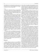
The patient was listed for insertion of a RVOT stent on day 12 of life due to worsening oxygen saturation requiring re-commencement of prostaglandin to keep baseline saturation above 70%. On this occasion, ini- tial right ventricle angiography was performed using a right femoral venous approach (Figure 2A and 2B). Similar to the third case, placement of a coronary wire across the tricuspid valve led to hypotension; there- fore, on this occasion, a decision was made to revert to a subxiphoid approach to minimize hemodynam- ic instability. Through a small subxiphoid incision, a 5-F sheath was placed through the anterior surface of the right ventricle (Figure 2C). Following sheath angi- ography, a 0.014” BMW wire was placed in the distal right pulmonary artery through a 4-F Terumo cathe- ter, and a 5 × 16-mm Formula 414 stent was deployed in a good position. Oxygen saturation increased to low 90%.
The patient had an uncomplicated post-opera- tive course and recovered well. Ten weeks following the initial procedure, weighing 3.14 kg, he under- went uncomplicated implantation of a further 6 × 20-mm Formula 414 stent via the right femoral vein for progressive cyanosis secondary to muscle hyper- trophy proximal to the RVOT stent. He subsequently underwent an uncomplicated full surgical repair at 5 months, weighing 5 kg.
Discussion
Despite signi cant advances in neonatal CPB, the mortality for symptomatic neonates with a diagnosis of ToF weighing < 2 kg has been reported as high as 49% [15]. This re ects the di culties of CPB in this age group, with infants having smaller circulating blood volumes, higher oxygen consumption rates, and highly reactive pulmonary vascular beds. In ad-
dition, infants have labile thermoregulation and im- mature organ systems with multiple implications for ischemic tolerance and in ammatory response [16]. However, despite these di culties, adopting a watch- and-grow approach has not been shown to improve outcomes [17, 18].
The unpredictability of pulmonary balloon valvu- loplasty combined with the high mortality of both modi ed BTT shunt placement and primary repair signaled the need for an alternative palliation op- tion—the RVOT stent. Since the initial report in 1997, numerous case series have reported on the use of RVOT stents in the palliation of ToF patients [4-10]. These mainly involved patients weighing > 2.5 kg.
In this series, we describe the evolution of our ap- proach to insertion of an RVOT stent in ToF patients weighing ≤ 2 kg. In four patients, we performed RVOT stenting to facilitate somatic and pulmonary artery growth to an adequate size suitable for primary re- pair. Previous reports demonstrate that mortality associated with early primary repair is highest in ne- onates with small pulmonary arteries (i.e., Nakata in- dex < 150 mm/m2) [19].
We have ne-tuned our approach to obviate the need for a long sheath to deliver the stent, which as- sists the approach in smaller infants; however, the po- tential hemodynamic instability seen with a sti cor- onary wire across the tricuspid valve may limit even this approach. Following the relative ease of stent delivery in the initial case, we felt that that a coronary stent could be delivered without much di culty. Lower pro le and shorter length stents appeared to course through the smaller heart more easily. How- ever, our second case highlighted the di culties that can arise with percutaneous stent insertion, partic- ularly ensuring adequate stent length without the availability of a long sheath, and the challenges with crossing a pre-existing stent in the RVOT. In the third case, hemodynamic instability required rapid stent deployment that ultimately led to stent migration. This case highlights the limited room for error and in- tolerance to stent manipulation within small hearts. In the fourth case, a hybrid perventricular approach via a subxiphoid incision was performed. This perven- tricular approach was previously described as a viable option in LBW neonates [10] and in this case allowed the infant to grow to successful neonatal repair.
Linnane N. et al.
RVOT Stenting in Small Infants
