
Page 35 - Journal of Structural Heart Disease Volume 4, Issue 2
P. 35
Meeting Abstracts
Journal of Structural Heart Disease, April 2018, Volume 4, Issue 2:56-65
DOI: https://doi.org/10.12945/j.jshd.2018.049.17
LAA CSI Focus Abstracts
LAA CSI Focus – November 17-18, 2017
THE VALUE OF 3D PRINTING FOR LAAO DEVICE SIZING
Vlad Ciobotaru1, Nicolas Combes2, Julien Ternacle3
1 Department of Cardiology, Hôpital Privé Les Franciscaines,
Nîmes, France,
2 Pediatric and Adult Congenital Heart Disease Department,
Clinique Pasteur, Toulouse, France
3 Department of Cardiology, CHU Henri Mondor, Créteil, France
Background: Percutaneous left atrial appendage occlusion (LAAo) is an alternative approach to medical therapy to prevent atrial bril- lation (AF) mediated stroke, in patients with a high stroke risk and contraindication for long-term oral anticoagulation. Despite an increasing rate of successful implantations, correct sizing and opti- mal device positioning remain a challenge. 3D printing has capability to create a highly accurate model of any structure and may be a use- ful approach for entire LAA anatomy, optimizing device size testing before LAAo..
Aim: To explore the usefulness of 3D printed LAA models to predict adequate sizing and risk of peri-device leaks or malposition post LAAo in a large patient cohort.
Published online: April 2018
Methods: 70 consecutive patients (CHA DS -Vasc score=4.5±1.5) with
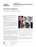
Figure 1. Panel A. Device-related thrombus (green arrow) in a patient with an o -axis prothesis leading to a cul-de- sac between the disc and the large pulmonary ridge. In ad- dition, note a gap between the disc and LA ostium (small arrow). Panels B, C, D, and E. Device predicted positions with regard to the sheath orientation in a printed mod- el. Panel B. O -axis device predicted by the printed model. Panel D. In vitro printed model superimposed on a cineangio- graphic view: improper alignment between LAA ostium axis with the sheath (red arrow). Panels C & E. Optimal device sealing using a the printed model and a larger size with an adequate sheath orientation (blue arrow)
pulmonary vein ridge and an inadequate transeptal site puncture. Complications rate was correlated with the mismatch between size used and printed model: odds ratio of 4.1 (1.5-12.7; p=0.01).
TM 22
a LAAo procedure with Amulet (St. Jude Medical, St Paul, MN, USA)
and a pre- and post-procedure cardiac CT, were included in three institutions between Feb 2014 and Mars 2017. A clinical follow-up was e ective for 17±10 months. Cavity segmentation was performed automatically based on CT Dicom data and manually adjusted to include the entire left atrium, then converted to a STL le and 3D printed with high resolution 50microns.
Results: Larges peri-device leaks >5mm occurred in 33% of patients. A mismatch between the model predicted size and the device used was a major predictor of peri-device leaks (AUC=0.85) with Predictive Positive Value (PPV)=82% and NPV=89% compared with CT sizing and TEE sizing (AUC=0.58 and 0.57, respectively p<0.001). Two cere- bral ischemic events occurred and four silent devices-related thrombi located in patients with an o -axis prosthesis. An o -axis position- ing was observed in 24% of patients. Predictive factors were a large
Fax +1 203 785 3346
E-Mail: jshd@scienceinternational.org http://structuralheartdisease.org/
© 2018 Journal of Structural Heart Disease Published by Science International Corp. ISSN 2326-4004
Accessible online at:
http://structuralheartdisease.org/
