
Page 36 - Journal of Structural Heart Disease Volume 4, Issue 2
P. 36
57
Meeting Abstracts
Conclusion: 3D-printed patient-speci c LA model allow more accu- rate sizing than TEE and CT measurements. It permits a pivotal train- ing, device testing and evaluation of optimal trans-septal puncture site with potentially important implications in minimizing proce- dure-related complications.
LEFT ATRIAL APPENDAGE CLOSURE WITH A NOVEL DEVICE: INITIAL EXPERIENCE AND MID-TERM FOLLOW-UP FROM A SINGLE CENTER
Paolo Pagnotta, Mauro Chiarito, Elton Pllah, Dennis Zavalloni Parenti, Marco Luciano Rossi, Riccardo Mantovani, Giulio Stefanini, Bernhard Reimers
Humanitas Clinical and Research Center, Interventional Cardiology Unit, Cardiovascular Department
Background: Left atrial appendage (LAA) closure is considered an e ective option in patients with non-valvular atrial brillation (NVAF) and contraindications to long-term oral anticoagulant (OAC) therapy. However, there are some concerns about safety of currently available devices.
Objective: Our aim is to provide an initial assessment on safety and e cacy of the novel LAA closure Ultraseal device in patients with NVAF and contraindications to long-term OAC therapy.
Methods: Thirteen consecutive patients with NVAF undergoing Ultraseal device implantation between July 2016 and April 2017 were included. All patients performed transesophageal echocardiography and computed tomography angiography prior to LAA closure.
Results: Procedural success was achieved in all patients except one who experienced incorrect device deployment but with complete LAA closure. Procedure duration halved from rst to last procedure performed. No adverse events, including pericardial e usion, were observed during index hospitalization. At mean follow-up (166±80 days) all patients were alive and free from major bleedings and isch- aemic strokes.
Conclusion: Our results suggest that the Ultraseal device is a safe and feasible option for LAA occlusion. Notably, the learning curve in this single-center experience was fast, paralleled by extremely low com- plication rates. These results should be considered hypothesis gener- ating and larger studies are mandatory.
ACUTE EMBOLISATION OF WATCHMAN PLUG ONTO AORTIC BIOPROSTHESIS FOLLOWED BY SUCCESSFUL PERCUTANEOUS REMOVAL
Konrad Pieszko1, Jarosław Hiczkiewicz2, Robert Sabiniewicz3, Wojciech Faron4, Sebastian Łukawiecki2, Dariusz Hiczkiewicz5 1 Szpital Nowa Sól; Oddział Kardiologii; Wsspzoz W Nowej Soli
2 Szpital Nowa Sól; Oddział Kardiologii; Ws Spzoz W Nowej Soli
3 Medical University of Gdansk; Interventional; Child Cardiology
4 Szpital W Nowej Soli; Oddział Kardiologii; Ws Spzoz W Nowej Soli 5 Szpital Nowa Sól; Oddział Kardiologii; Ws Spzoz Nowa Sól
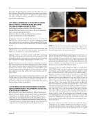
Figure 1. Panel A. Watchmann Device placed in LAA. Panel B. Con rmation of correct placement in TEE. Panel C. Device em- bolising aortic bioprosthesis. Panel D. Doppler showing residual ow through the device during reanimation.
History, Physical and Indication For Intervention: Our patient was an 80-year old woman who underwent implantation of aortic biopros- thesis (Shellhigh 27) 8 years earlier due to severe aortic stenosis. She su ered from chronic kidney disease (stage G3a), hypertension and paroxysmal atrial brillation, but no coronary artery disease. She had history of multiple severe bleedings from the lower digestive tract during oral anticoagulation with with rivaroxaban.
Intervention: The procedure initially went with no complications using sedative drugs only and no general anesthesia. Patients’ LAA had “chicken wing” morphology and the maximum width of ostium was 14,1mm measured in TEE during the procedure. Initially a Watchman 27mm was used, but it had to be switched to a Watchman 21mm because of excessive protrusion. Correct localization of the Watchman device was con rmed in scopy and TEE, as shown on (Fig.1, panel A and B, respectively) as well as by tug test with 9% device compression (19/21mm). Color doppler showed no signi cant peri-device ow. A few minutes after the procedure was nished, while the patient was still in the cath lab, there was a cardiac arrest with pulseless electrical activity. Immediately we begun resuscitation and reintroduced the TEE probe. The echo image showed embolisa- tion of the plug on the aortic bioprosthesis with almost complete obstruction of ow as shown on (Fig 1. panel C). During heart mas- sage only a very small jet of ow across the plug was observed as shown on (Fig 1. panel D). Resuscitation was continued according to the ERC guidelines. Six minutes after arrest VF was observed and patient was de brillated once with 200J successfully. Position of the device and lack of su cient ow through aortic valve required imme- diate intervention.
Meanwhile, a EN Snare vascular loop 6x10 was introduced using a 11F Cordis vascular sca old in left femoral artery. The loop was pro- truded through bioprosthesis valves and we succeeded in catching the device and moving it down to the abdominal aorta. Soon after
Hijazi, Z
2017 LAA CSI Focus Abstracts
