
Page 39 - Journal of Structural Heart Disease Volume 4, Issue 2
P. 39
Meeting Abstracts
60
Conclusion: The image-fusion techniques allow combining comple- mentary information for improving the visualization of the patterns of interest and the procedure’s planning and execution. By image-fu- sion is possible to take advantage of both the higher temporal reso- lution of the echocardiographic images and the greater anatomical accuracy of the CT models. Moreover, the use of 3D models in the registration procedure allows exploiting them for the LAA anatom- ical and functional characterization, for biomechanical simulation and for 3D printing in support of pre-operative planning. The mul- timodal-image-fusion overcomes the limitations of the standard image-techniques routinely used in cathlab.
SIMULTANEOUS PERCUTANEOUS CLOSURE OF LEFT ATRIAL APPENDAGE AND PATENT FORAMEN OVALE Costantino R. Costantini, Sergio G Tarbine, Costanitno O. Costantini, Admar de Souza, Daniel Zanuttini, Marcelo Freitas Santos, Marcos Denk
Hospital Fundacao Francisco Costantini; Cardiology Department; Interventional Cardiology
History: L.J.R, male, 69 yo, admitted with history of dyslipidemia, inferior limb venous insu ciency & several atrial brilation episodes. Echocardiography showed no thrombus in left atrium, left atrial appendage (LAA) with favorable anatomy to closure, and the pres- ence of patent foramen ovale (PFO) with right to left shunt detected by saline microbubbles.
Images:
Indication With CHADS and CHA2DS2-VASC scores 1, oral anticoagu- laion become among the indications. Because of clinical reasons and patient preference, percutaneous closure of the LAA and the PFO was performed.
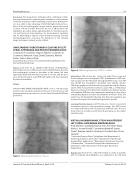
Figure 1. Delivering Watchman 27 device. Journal of Structural Heart Disease, April 2018
Figure 2. Delivering Amplatzer 25 PFO occluder.
Intervention: LAA closure was carried out under uoroscopy and Transesophageal echocardiography (TEE). A multipurpose (MP) cath- eter was placed in the left atrium through the PFO. Using a 0,35 sti wire, the MP was changed for a dedicated 14 Fr delivery catheter. LAA Angiography was performed using a pigtail through de delivery system. After measurements by uoroscopy & TEE, a 27 Watchman device was released closing the LAA. Using the same delivery system, a 25 amplatzer PFO device was implanted con rming its size, position and stability. Doppler and saline microbubbles showed no shunt. The patient was discharged the following day. Oral anticoagulation was used and changed to ASA and Clopidogrel after 6 months.
Learning Point-Conclusion: LAA Percutaneous closure is at present a treatment option in stroke prevention settings. Also PFO closure is recommended when risk of paradoxical embolism exist. The case shows that it is possible to perform both percutaneous procedures simultaneously.
VIRTUAL HAEMODYNAMIC STUDY ON DIFFERENT LEFT ATRIAL APPENDAGE MORPHOLOGIES
Andy Olivares1, Guadalupe Garcia-Isla1, Etel Silva2, Marta Nuñez1, Jerome Noailly1, Constantine Butako 1, Xavier Freixa3, Damian Sanchez-Quintana4, Tom de Potter2, Oscar Camara5
1 Universitat Pompeu Fabra; 2 Arrhythmia Unit, Department of Cardiology, Cardiovascular Center; 3 Department of Cardiology, Hospital Clínic de Barcelona; 4 Department of Anatomy, University of Extremadura; 5 Universitat Pompeu Fabra; Department of Information and Communication Technologies; Biomedical Engineering
Background: Around 90% of thrombi leading to stroke in patients with non-valvular atrial brillation are originated in the left atrial
Volume 4, Issue 2:56-65
