
Page 41 - Journal of Structural Heart Disease Volume 4, Issue 2
P. 41
Meeting Abstracts
62
RAO 30, CRAN 15 – 23.4 mm; RAO 30, CAUD 15 – 24.2 mm. Amplatzer Cardiac Plug 26 mm was selected for implantation. LAAO implanta- tion was successfully performed. Traction test was negative. LAAO position stable. The second day after LAAO implantation trans-esoph- ageal Echo was performed and revealed no abnormalities. The patient was discharged on the 3-d day after the procedure. Rivaroxaban was prescribed for 3 months. At 3-month follow–up TEE was performed. The Patient had dislodgement of a disk-part of the occluder into dis- tal direction of the appendage. The upper part of the disk slept under the ridge. Pannus formation is currently registered above the disk at previous disk position, so that it was covering the main part of the ridge. We could describe triangle formation which is formed by the disk surface, pannus and the ridge (Pic. 1). The patient was switched to warfarin aiming INR about 2,5-3,0. At 6-month follow–up TEE was performed. This diagnostic revealed thrombus formation on LAAO disk surface in size of 1,46*0,41cm (Pic. 2). The pannus was not visu- alized anymore. Warfarin dose was increased, aiming INR about 3,0. At 9-month follow-up no more thrombotic masses were veri ed. The patient was recommended to continue intake of Rivaroxaban.
Learning Points of the Procedure: Pannus formation is a rare addi- tional complication phenomenon after LAA occluder disk dislodge- ment. Pannus is a substrate for further thrombus development. In order to prevent LAAO dislocation it is important to choose the right size of the device, to be sure not to put the device too deep into LAA. Achieve stable position of the occluder is always import- ant. Adequate anticoagulation after LAAO implantation should not be underestimated.
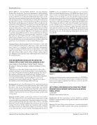
RT3DTEE con be very helpful for the procedure. A case of previous ASD closure with Amplatzer device n.22 (PANEL B) in which is possi- ble to see the close relationship between the device, the aortic root and the roof of LA. Clearly to access the LA without puncturing the transseptal device is possible only in the inferior part of IAS just below the device through the adjacent native septum. 3DTEE imaging facili- tates the procedure by providing in one single view detailed pictures of the targets in relation to each other. Congenital corrected trans- position (cGTA) in situs viscerum inversus is a rare cardiac malforma- tion characterized by the combination of discordant atrio-ventricular and ventriculo-arterial connections (PANEL C). The great arteries are parallel to each other. By far the most important structure to avoid puncturing is in our case the pulmonary trunk due to its close posi- tion with IAS.
Figure 1.
Conclusions: Performing transseptal puncture the role of RT3DTEE is an added value showing anatomical images in multiple perspective, even more if complex congenital heart disease or previous percuta- neous repair are present.
LEFT ATRIAL APPENDAGE OCCLUSION FOR STROKE PREVENTION IN PATIENTS WITH VALVULAR ATRIAL FIBRILLATION
Joelle Kefer
Cliniques Universitaires Saint-Luc
Background: Left atrial appendage occlusion (LAAO) is a valuable therapy for stroke prevention among patients with non valvular atrial brillation (NVAF).
Objective: The aim of the present study was to evaluate the feasibil- ity and the safety of LAAO in patients with valvular atrial brillation ( VAF).
THE INTRAOPERATIVE ROLE OF RT3DTEE FOR TRANSSEPTAL PUNCTURE: CHALLENGING CASES Laura Lanzoni1, Giulio Molon2, Guido Canali3, Stefano Bonapace4, Clementina Dugo1, Andrea Chiampan5, Enrico Barbieri6
1 Ospedale Sacro-Cuore Don Calabria; Ospedale Sacro-Cuore Don Calabria; Ospedale Sacro-Cuore Don Calabria, 2 Cardiology Division, Sacro Cuore Hospital; Interventional; Adult Cardiology, 3 Ist. S.Cuore Don Calabria; Interventional; Adult Cardiology, 4 Ospedale Sacro- Cuore Don Calabria; Ospedale Sacro Cuore- Don Calabria; Ospedale Sacro Cuore- Don Calabria, 5 Cardiology Division, Sacro Cuore Hospital; Non Invasive; Adult Cardiology, 6 Ospedale Sacro-Cuore Don Calabria; Invasive Cardiology; Adult Cardiology
Background: Access to the left atrium (LA) for several interventional procedures is obtained by transseptal puncture that permits direct passage to the LA via the interatrial septum (IAS) and systemic venous system. Careful knowledge of anatomy of IAS and its anatom- ical variations (thickened, aneurysmal, previous procedures, presence of PFO) is important for the success of the procedure and the safety of the patient.
Methods: RT3DTEE permits good images of IAS and correct evalua- tion of the distance from aorta and left atrium walls; may be useful in patients with unusual anatomy of interatrial septum such as lipo- matous hypertrophy (LH) and very small surface of fossa ovalis (FO) (Panel A) to reach exactly the right point to puncture and to see the tenting of the fossa by the catheter tip. In patients with congenital heart disease who have undergone surgical or percutaneous repair, access from the RA to the LA could be challenging and the use of
Journal of Structural Heart Disease, April 2018
Volume 4, Issue 2:56-65
