
Page 15 - Journal of Structural Heart Disease Volume 4, Issue 3
P. 15
Original Scienti c Article
72
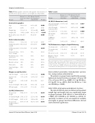
Table 1. Demographic and echocardiographic characteristics of patients in two groups (short vs. prolonged procedure time).
Table 1 (cont.).
LA, MV, LV dimensions (cont.)
Groups
Group I ( n = 56) Short proce- dure time
Group II (n=25) Prolonged pro- cedure time
P-value
Groups
Group I ( n = 56) Short procedure time
Group II (n=25) Prolonged pro- cedure time
P-value
General demographics
LA (Lateral) length, indexed to BSA
LA (A-P) length, indexed to BSA
LA volume cm3
MV annulus (Z-score)
LVEDD size (Z-Score)
LV SF
33.16 ± 7.48
26.95 ± 7.3
19.9 ± 10.4 −0.02 ± 0.81
−1.2 ± 1.5 33.63 ± 3.56
38.08 ± 9.89 32.67 ± 9.59
14.8 ± 10.6 −0.18 ± 1
−0.86 ± 1.54 35.38 ± 5
0.017
0.005
0.050
0.476 0.363 0.081
0.001
0.528
0.198 0.016
Age
Male
Weight (kg)
Height (cm)
Body surface area (m2)
6.98 ± 3.92 41.8%
21.9 ± 11.8 114.62 ± 20.04 0.8 ± 0.3
4.47 ± 3.86 41.7%
15.14 ± 8.06 96.92 ± 16.77 0.6 ± 0.25
12.17 ± 3.91 0.93 ± 0.37
0.13 ± 0.04
16%
8.3%
12.5% 8.3% 37% 4.2%
11.66 ± 2.91 12.46 ± 4.27
10.9 ± 3.82
5.55 ± 1.94 11.2 ± 3.4
31.71 ± 6.8
22.33 ± 5.63 19.04 ± 4.54 54.25 ± 14.18
0.009
0.990
0.013 < 0.001 0.005
0.706
<0.001
0.014
0.994
0.629
0.838 0.164 0.065 0.910
0.050
0.426 0.211
0.290 0.339
0.015 0.014
0.160
0.013
Defect related variables
ASD size
ASD size/patient’s weight
ASD size/patient’s length
Aneurysm devia- tion > 10 mm
Prominent eusta- chian valve
Chiari network
Double contour
Flimsy rims
Septal malalign- ment
11.8 ± 0.64 ±
0.10 ±
16.1%
5.5%
10.9% 1.8% 18% 3.6%
3.99 0.28
0.03
TV, RV dimensions, degree of septal attening
SVC rim length
Superior rim length
AV valve rim length
Aortic rim length IVC rim length
13.13 ± 2.98 11.73 ± 3.43
12.35 ± 5.06
6.09 ± 2.08 12.19 ± 4.27
LA, MV, LV dimensions
LA (coronal) length
LA (lateral) length LA (A-P) length
LA (coronal) length, indexed to BSA
35.82 ± 6.77
25.62 ± 5.25 20.47 ± 3.92 46.73 ± 11.05
TV annulus size
TV annulus size ( Z-score)
RV size ( Z-score)
Degree of septal attening ≥ 2
24.67 ± 3.32 1.14 ± 0.59
8.94 ± 1.59
16.4% 41.7%
22 ± 3.28 1.04 ± 0.83
8.38 ± 2.05
Margins around the defect
Data are presented as mean ± standard deviation or as number (percentage) of patients. ASD = atrial septal defect. SVC = superior vena cava. AV = atrioven- tricular valve. IVC = inferior vena cava. LA = left Atrium. A-P = anterior-poste- rior. MV = mitral valve. LVEDD = left ventricular end diastolic dimension. LVSF = left ventricle shortening fraction. TV = tricuspid valve; RV = right ventricle.
device-related parameters, hemodynamic parame- ters, and procedure-related details.
The patients in group II were signi cantly younger, lighter and shorter than those in group I: (4.5 years SD 3.9 vs. 6.9 years SD 3.9; p = 0.009), (15.1 kg SD 8.06 vs. 21.9 kg SD 11.8; p = 0.013), (96.9 cm SD 16.8 vs. 114.6 cm SD 20.04; p < 0.001).
Defect (ASD), atrial septum and adjacent structures
The ratio of ASD size (mm) in relation to the patient weight (kg) and length (cm) was signi cantly larger in group II patients: (0.93 SD 0.37 vs. 0.64 SD 0.28; p < 0.001), (0.13 SD 0.04 vs. 0.1 SD 0.03; p = 0.014), re- spectively. Incidence of all non-numerical variables was higher in group II but these di erences did not reach statistical signi cance.
Journal of Structural Heart Disease, June 2018
Volume 4, Issue 3:69-78
