
Page 16 - Journal of Structural Heart Disease Volume 4, Issue 3
P. 16
73
Original Scienti c Article
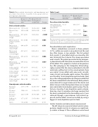
Table 2. Device-related characteristics and hemodynamic and procedural characteristics of patients in two groups (short vs. prolonged procedure time).
Table 2 (cont.).
Procedure-related variables
Groups
Group I ( n = 56) Short proce- dure time
Group II (n=25) Prolonged pro- cedure time
P-value
Groups
Group I ( n = 56) Short procedure time
Group II (n=25) Prolonged pro- cedure time
P-value
Device related variables
Time between de- vice deployment & release(min)
Procedure time (min)
Fluoroscopy time (min)
6 ± 2.1
49.8 ± 18.13 10.92 ± 6.71
40.72 ± 57 < 0.001
93.32 ± 45.4 < 0.001
25.92 ± 15.36 < 0.001
Device waist size
Device LA disc size
Device waist di- ameter / patient’s weight
Device waist di- ameter / patient’s height
Device waist diameter/total septal length
Device waist diameter/LA coronal length
Device waist di- ameter/LA lateral length
Device waist diameter/LA (A-P) length
LA disc size/LA coronal length
LA disc size/LA Lateral length
LA disc size/LA (A-P) length
LA disc size/total septal length
13.13 ± 4.21 26.32 ± 4.86
0.71 ± 0.31
0.12 ± 0.04
0.44 ± 0.56
0.37 ± 0.11
0.53 ± 0.18
0.66 ± 0.22
0.75 ± 0.16 1.07 ± 0.28 1.33 ± 0.32 0.88 ± 1.06
14.28 ± 4.89 25.06 ± 6.46
1.04 ± 0.44
0.15 ± 0.05
0.41 ± 0.11
0.46 ± 0.14
0.67 ± 0.23
0.77 ± 0.25
0.81 ± 0.2 1.19 ± 0.42 1.38 ± 0.45 0.72 ± 0.19
32.48 ± 9.87 19.78 ± 6.79 26.39 ± 8.06 11.65 ± 4.09 1.8 ± 0.71
0.281
0.334
< 0.001
0.003
0.791
0.003
0.006
0.039
0.218 0.134 0.580 0.469
0.027
0.008 0.186 0.037 0.733
Data are presented as mean ± standard deviation or as number (percentage) of patients. LA = left atrium. A-P = anterior-posterior. RV = right ventricle. PA = pulmonary artery PA. Qp:Qs = pulmonary ow : systemic ow.
Procedure failures and complications
Device embolization occurred in three patients (3.7%), within one week in one patient and the next day, after closure, in two patients. The rst patient had a central 11 mm defect which was closed by 10.5 mm Occlutech, Flex II device. The closure procedure went smooth. The patient presented to the emergen- cy department with chest pain one week after closure of the defect. The symptoms started on the previous day after jumping on the trampoline. Echocardiog- raphy revealed that the device embolized to the left ventricular out ow tract. In the second patient, there was central 12 mm defect with some aortic rim de - ciency (5 mm) and double septal contour. The defect was closed by 12 mm Amplatzer septal occluder. Next day, echocardiography before discharge revealed that the device embolized to the left atrium. Both patients were referred for surgical removal of the device and defect closure. In the third patient, there was an eight mm central defect and double septal contour. The de- fect was closed by 9 mm Amplatzer septal occluder. Next day, chest X-ray before discharge revealed that the device embolized to the descending aorta. It was retrieved through a percutaneous trans-arterial ap- proach and the defect closed successfully by using a bigger (12 mm) Amplatzer septal occluder.
Cardiac chambers and valve annulus measurement
The left atrium (i) coronal, (ii) lateral and (iii) antero- posterior lengths and (iv) its volume was signi cant- ly smaller in Group II. These di erences were found
Hemodynamic-related variables
RV systolic pressure
PA mean pressure
PA Systolic pressure
PA Diastolic pressure
Qp:Qs
27.91 ± 7.38 16.48 ± 3.9 24.27 ± 5.65 9.77 ± 3.37 1.74 ± 0.68
El-Segaier M. et al.
Predictors for Prolonged ASD Closure
