
Page 23 - Journal of Structural Heart Disease Volume 4, Issue 3
P. 23
Original Scienti c Article 80
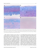
Figure 1. Pulmonary artery histology from a patient with Pulmonary artery aneurysm. Panel A. Trichrome/elastin stain demonstrating severe elastic ber loss (40x) consistent with grade 3 arteriopathy (Niwa et al, Circulation 2001). Panel B. At high power, there is dis- ruption (arrow) and fragmentation of the remaining elastic bers (trichrome/elastin, 100x). Panel C. The alcian blue stain highlights translamellar mucoid extracellular matrix accumulation (100x). Panel D. The extracellular mucoid matrix materials (glycosaminogly- cans) are digested following treatment with hyaluronidase (100x).
When pulmonary aneurysms do occur, they are usually secondary to a variety of factors, most com- monly pulmonary arterial hypertension and/or con- genital cardiac shunts. Less commonly pulmonary artery aneurysms may be secondary to infection (syphilis and tuberculosis), congenital arteriopathy (e.g. Marfan and Turner syndrome), auto-immune conditions (Behcet’s) or congenital pulmonary valve abnormalities (Tetralogy of Fallot with absent pulmo- nary valve, pulmonary stenosis and/or regurgitation) [2]. Pulmonary artery dilation may be present in pa- tients with pulmonary valve stenosis, however, cata- strophic complications such as dissection or rupture
are rare (Roberts WC et al. AJC 2017, Adodo et al. Ann Thorac Surg 2017, Koretzky Circulation 1969). The his- topathologic characteristics are similar to those seen in the aortas of those with congenital aortic valve dis- ease [9]. The association between aortic dilation and congenital aortic stenosis is well known, however, the degree of aortic dilation is not directly related to the degree of valvular stenosis. This suggests a congeni- tal etiology to the aortopathy in bicuspid aortic valve patients that may be independent of valvular hemo- dynamics. Could a similar logic exist for congenital pulmonary valve stenosis? The de nition of an aneu- rysm is “focal dilation of a blood vessel involving all
Journal of Structural Heart Disease, June 2018
Volume 4, Issue 3:79-84
