
Page 49 - Journal of Structural Heart Disease Volume 4, Issue 3
P. 49
Meeting Abstracts 106
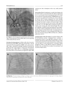
29. Figure 1. Hand Angiogram in LAO Cranial view from the right carotid showing a Vertical PDA (white arrow) supplying small sized pulmonary arteries.
Indication for Intervention: In infants with critical cyanotic heart disease, like HRHS, pulmonary blood ow is ductal dependent. The management of HRHS can be done in mul- tiple stages of palliation and PDA stenting is the preferred method of rst stage palliation compared to the Modi ed Blalock-Taussig Shunt (BTS) in establishing pulmonary blood ow. Due to the limited prostaglandin supply, PDA
stenting was thus attempted in this very small preterm infant.
Intervention: Ductal stenting via a carotid arterial approach was done on the second day of life. The right carotid artery was accessed under direct visualization and a 4F sheath was inserted in standard technique. A BMW 0.014 inch cor- onary wire was guided in the pulmonary arteries through the ductus after an initial angiogram was done through the sheath side port. The ductal length was measured and an Omega bare metal coronary stent measuring 2.75mm x 12mm was inserted over the wire and was subsequently positioned across the PDA without jailing the pulmonary arteries and covering the entire ductal length. The stent was in ated to rated burst pressure giving an inner diame- ter of 3mm. A repeat angiogram was done in order to doc- ument adequate stent position and to visualize pulmonary blood ow. Transthoracic echocardiogram after the proce- dure documented adequate PDA stent ow into con uent pulmonary arteries.
Learning Points of the Procedure: In preterm infants with life threatening cyanotic congenital heart condition, a management option is through PDA stenting in order to maintain and ensure pulmonary blood ow. The procedure is limited by patient size and prematurity but with proper care and skill, it can be performed in a premature infant weighing as little as 1.2kg. Ductus arteriosus stenting is a feasible, safe, and e ective procedure for premature and low birth weight infants.
AB
29. Figure 2. AP view showing an Omega bare metal coronary stent in place (Panel A); Hand injection angiogram showing adequate stent position supplying both pulmonary arteries (Panel B).
Journal of Structural Heart Disease, April 2018 Volume 4, Issue 2:85-113
