
Page 50 - Journal of Structural Heart Disease Volume 4, Issue 3
P. 50
107
Meeting Abstracts
30. TRANSCATHETER INTERVENTION FOR PARAVALVULAR LEAK IN MITROFLOW BIOPROSTHETIC PULMONARY VALVE Vishal Kaley, E. Oliver Aregullin1, Bennett Samuel, Joseph Vettukattil
Spectrum Health Helen Devos Children's Hospital; Congenital Heart Center; Congenital Heart Disease
History and Physical: Paravalvular leak (PVL) is a compli- cation due to suture dehiscence between the sewing ring and native tissue resulting in regurgitation around the replaced valve. The standard treatment for pulmonary PVL is surgical repair or valve replacement. However, sur- gery is associated with greater morbidity and mortality. Transcatheter intervention for aortic and mitral valve PVL is e ective and known to have better long-term outcomes than surgery, which has a 12-year survival of 30-40% and high rate of recurrence. In the setting of pulmonary PVL, transcatheter approach may be a useful technique with optimal outcomes.
A 22-year-old male with tetralogy of Fallot and bilateral peripheral pulmonary artery (PA) stenosis presented with multiple episodes of syncope, dyspnea on exertion (NYHA class III) and worsening lower extremity edema. He had a transannular patch repair early in life. Due to severe pulmo- nary regurgitation (PR), his pulmonary valve was replaced with a 27 mm Mosaic tissue valve at 8 years of age. He was noted to have free PR, and depressed systolic function at 15 years of age. Subsequently, he underwent pulmonary valve replacement with a 25mm Mitro ow bioprosthetic valve and intraoperative stenting of the branch PAs. Post- operative period required extracorporeal membrane
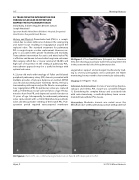
30. Figure 1. CT on True3D Viewer (Echopixel, Inc., Mountain View, CA) showing paravalvular tunnel measuring 8x6x9 mm in the posteromedial side of the pulmonary valve.
oxygenation support and prolonged tracheostomy lead- ing to severe post-traumatic stress syndrome. On Holter monitoring, he was noted to have ventricular tachycardia.
Imaging: (See Figures 1 & 2).
Indication for Intervention: In view of worsening dyspnea, syncope, and edema, PVL closure was considered (Figure 1). Considering his complex history and associated risks with redo-sternotomy, a multi-disciplinary team recom- mended transcatheter PVL closure.
Intervention: Moderate stenosis was noted across the Mitro ow valve at the pulmonary position with gradient of
30. Figure 2. Panel A. PVL size con rmed using Armada balloon; Panel B. AVP II deployed in the PVL.
Hijazi, Z 2017 CSI Africa Abstracts
