
Page 14 - Journal of Structural Heart Disease Volume 4, Issue 5
P. 14
209
New Technology
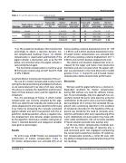
Table 1. VO and CT-aided motion compensation results.
Valve type
Pigtail catheter cusp position
Number of cases
Frames with activated MC (%)
VO MC
Relative displacement error (mm)
Absolute displacement error (mm)
Frames with activated MC (%)
CT-aided MC
Relative displacement error (mm)
Absolute displacement error (mm)
CoreValve Lowest 8
82
87
89
84
-1.10±2.61
0.09±2.56
-1.24±2.72
-1.09±2.65
2.00 85
1.97 98
2.71 80
2.24 84
-1.13±2.91 2.15
-0.15±2.45 1.50
-0.09±3.02 2.48
-0.77±2.92 2.22
Sapien
Lowest 4
Middle 12
Total - 24
1f-g). The anatomical roadmap is then transformed accordingly to obtain a real-time dynamic mo- tion-compensated roadmap (Figure 1h). Motion compensation is deactivated automatically if the pigtail catheter is obstructed, such as by the TEE probe, and activated when the pigtail catheter is successfully found again.
The live motion compensation is real-time up to 30 frames per second using an Intel® Xeon E5-1620 v3 CPU 3.50GHz.
Automatic Motion Compensation Evaluation Protocol
The use of a motion compensated overlay occurs during the device positioning and deployment phase, so we post-processed X-ray data of 24 cases during this phase to evaluate the algorithmic performance. None of these datasets were used for algorithm de- velopment.
First, the percentage of frames in which motion compensation was correctly activated by the algo- rithm was determined. Secondly, the relative and ab- solute displacement error were determined for every X-ray frame by comparing the manually annotated pigtail catheter and aortic root position with the al- gorithmic roadmap position, where a negative rela- tive displacement error denotes deeper positioning by the algorithm. Continuous variables were given as mean ± standard deviation and categorical variables were given as percentages.
Results
For all 24 cases (25,607 frames) we evaluated the per formance of motion compensation ( Table 1). VO motion compensation was activated 84% of all
frames yielding a relative displacement error of -1.09 ± 2.65mm and 2.24mm absolute displacement error. CT-aided motion compensation was activated 84% of all frames yielding a relative displacement -0.77 ± 2.92mm and 2.22mm absolute displacement error.
The relative and absolute displacement error in- creased for the larger and hence more obstructive CoreValve and also increased when the pigtail cath- eter was positioned in the more obstructive middle position (Table 1). Overall VO and CT-aided motion compensation demonstrated similar performance.
Discussion
We have used the pigtail catheter as a contrast-in- dependent landmark for motion compensation during TAVI without any need for software interac- tion. To our knowledge, only one approach has suc- cessfully tracked the aortic valve plane by using the calcifications on the aortic valve as contrast-indepen- dent landmarks [8]. A clinical trial correlated this ap- proach with a promising reduction in the incidence of conduction disorders [9]. The feasibility of the ap- proach was limited by the need to manually anno- tate the calcifications after every repositioning of the C-arm. Additionally, not every patient may have suffi- cient visible calcifications [10]. All currently available CT fusion solutions provide static overlays only.
Of the two motion compensation methods eval- uated: VO has the advantage of requiring only a well-contrasted aortic root angiogram representing the current aortic anatomical situation. CT-aided mo- tion compensation provides a richer 3D view, with the ability to integrate pre-procedural planning in the live roadmap.
Assink N. et al.
Automated Motion Compensation for TAVI
