
Page 35 - Journal of Structural Heart Disease Volume 4, Issue 5
P. 35
Case Report
230
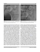
Figure 3. Balloon pulled out of the groin. The balloon, mostly within the 8 Fr sheath, has been pulled out of the left groin so that it could be exposed and popped manually.
described in the early literature for the balloon atrial septostomy procedure [2-6]. In some cases, successful deflation of the balloon was eventually achieved by use of the stiff end of a wire advanced either through the second lumen of the balloon catheter or through a second end-hole catheter. With the wire held in po- sition just past the tip of the catheter, the balloon was pulled back onto the wire in order to puncture the balloon [2,5]. In other cases, the balloon was punc- tured by a fine needle introduced percutaneously, either transhepatically or through the chest wall [3,4]. Both of these approaches have significant risks as- sociated with them. With the first approach, there is the risk of vessel/cardiac damage from the wire. With the latter, there are obvious risks through transcuta- neous needle access of the balloon of damage to the heart and surrounding structures. According to Hijazi et al., when a balloon septostomy catheter does not deflate, the first thing to do is to pass a guide wire in the balloon lumen to clear any obstruction. This ma- neuver was not performed in our case. If this does not work, an injector should be connected to the balloon and 3-5 cc of contrast injected under pressure using 300 psi in order to rupture the balloon [7]. With this
Figure 4. Deflated balloon within sheath. After the balloon has been manually deflated, it has now been pulled back into 4 Fr RIJ sheath so that it can safely be removed from the body.
approach, rupture of the balloon at high pressure has the risk of embolization of the balloon fragments[8]. Finally, if this does not work, one can try using the stiff end of a guide wire through a second catheter [7].
In order to test the technique of balloon rupture, we overinflated a series of balloon catheters ex vivo to see at what pressure the balloons burst and how they tore. First, we manually burst a series of balloons by slowly over-inflating the balloon with a BasixTouch inflation device (Merit Medical, South Jordan, UT) un- til the balloon burst. It should be noted that balloon rupture mechanisms are subject to variations due to balloon materials system fatigue. Table 1 lists the sizes and types of balloons tested as well as the burst pres- sures and type of hole or tear created. During manual over-inflation, all Sterling balloons as well as the Opta Pro, Palmaz Blue and Valeo balloons had longitudinal tears. The Dorado Balloon burst with a pinhole tear in the proximal balloon. The Atlas Gold Balloon did not burst, but the high pressure created a connection be- tween the wire lumen and the balloon lumen within the catheter. This balloon remained unable to deflate. We additionally burst a Miller-Edwards Balloon Sep- tostomy Catheter (Edwards Life sciences, Irvine, CA).
Journal of Structural Heart Disease, October 2018
Volume 4, Issue 5:228-233
