
Page 37 - Journal of Structural Heart Disease Volume 4, Issue 5
P. 37
Case Report
232
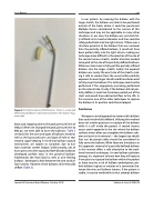
Figure 5. Complete rupture of BAS balloon. Shown is a ruptured Miller-Edwards Balloon Septostomy Catheter with balloon frag- ment seen.
kept auto-stopping due to the peak pressure limit of 300 psi. When we changed the peak pressure limit to 600 psi, we were able to burst the balloons. Table 2 similarly lists the sizes and types of balloons tested as well as the burst pressures and type of hole or tear created, again keeping in mind that balloon rupture mechanisms are subject to variations due to bal- loon materials system fatigue. Unfortunately, not all burst pressures were captured. Both Dorado balloons developed pinhole tears in the proximal balloon. Additionally, the Atlas Gold as well, as one Sterling balloon, developed a hole between the wire and bal- loon lumens. However, these balloons did eventually deflate (Table 2).
In our patient, by covering the balloon with the larger sheath, the balloon was able to be positioned outside of the body where it could be punctured. Multiple factors contributed to the success of this technique and may not be applicable in many other situations. In our case, the balloon was not stuck ful- ly inflated to its maximal diameter and thus could be safely pulled back into the right atrium. If there was a structure proximal to the balloon that was narrower than the partially inflated balloon, it could not have been pulled safely into the right atrium, making our technique more difficult. In the selection of the size of the second venous sheath, careful attention needed to be paid to the size of the partially deflated balloon. While our intent was to fully pull the partially inflated balloon into the larger sheath, luckily the exposed balloon was nearly the exact size of the sheath, mak- ing it safe to remove from the vessel orifice partially exposed. An even larger sheath could have been used to fully cover the balloon. This technique could not be performed if the angioplasty was being performed on the arterial side. Finally, if the balloon did not par- tially deflate, it could not have been pulled out of the stent and would have obstructed flow to the LPA. In this scenario, one of the other techniques to rupture the balloon in its position could be employed.
Conclusion
We report a novel approach to removal of a balloon that could not be fully deflated. Utilizing this method does not involve puncture or rupture of the balloon while it is still inside the patient. A second venous access point opposite to the site where the balloon catheter enters often can straighten the balloon cath- eter and assist in its retrieval — the largest size sheath that can be placed safely should be considered to fully cover the balloon. While there are limitations to this approach, removal of a partially deflated balloon in this manner offers a safe alternative to the tradi- tional removal techniques. As new balloon catheters emerge, it is important to know how they will rupture if one plans to rupture the balloon within the patient as there may be a risk of balloon embolization, pin- hole balloon rupture or creation of a connection be- tween the wire and balloon lumens. If the patient is stable, it may be worthwhile to first attempt balloon
Journal of Structural Heart Disease, October 2018
Volume 4, Issue 5:228-233
