
Page 40 - Journal of Structural Heart Disease Volume 4, Issue 5
P. 40
235 Case Report
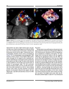
Figure 1. Panel A. TEE showing severe mitral regurgitation through the AML perforation. Panel B. 3D-TEE showing the wire passing from the AV to AML perforation. Panel C. Fluoroscopic view showing the complete arteriovenous loop and delivery sheath introduc- tion. Panel D. 2D-TEE view during passing the mechanical aortic valve with the wire, showing moderate aorticregurgitation(left); after device implantation and removal of the wires, there is no aortic regurgitation (right).
replacement was done. Eight months later, she pre- sented to our center with progressive SOB with NYHA class III-IV. Cardiovascular examination revealed 4/6 holosystolic murmur at the apex. Transthoracic echo- cardiography revealed severe mitral incompetence (Figure 1A). Transesophageal echocardiography (TEE) showed 5x5mm anterior mitral leaflet (AML) perfo- ration through the A2 segment with moderate pul- monary hypertension (estimated systolic pulmonary artery pressure of 50 mmHg). The aortic valve showed a mean gradient of 18 mm/Hg across the AV with no valvular or paravalvular leaks. The left ventricular ejection fraction (LVEF) was 55% and the left ventric- ular end systolic diameter was 46 mm. Several blood cultures were taken and they showed no bacterial growth. As the patient refused redo surgery, she was referred for a possible percutaneous closure of AML perforation.
Procedure
The procedure was performed under general anes- thesia with three-dimensional TEE guidance (PHILLIPS iE33 Cardiovascular Ultrasound, USA) and periproce- dural prophylactic antibiotics were given. The chal- lenges were crossing the defect in the A2 segment, selecting the appropriate device and the AML be- havior after device deployment. Very low transseptal puncture was intended to create a straight tract with- out tension on the AML during closure. We anticipat- ed that crossing the defect from the LA side will be ex- tremely difficult due to leaflet’s movement away from and parallel to the crossing wire with each heartbeat. In addition, crossing through the mechanical aortic valve may carry the challenge of hemodynamic insta- bility or mechanical disruption of the valve. Arterial and venous femoral accesses were secured and hep- arin was given. Transseptal access was done; tip de- flectable catheter (Agilis St Jude) 8.5 F was introduced
Abuelatta R. et al.
Transcatheter Repair of AML Perforation
