
Page 41 - Journal of Structural Heart Disease Volume 4, Issue 5
P. 41
Case Report 236
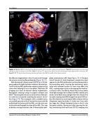
Figure 2. Panel A. 3D-TEE showing the device located within the AML sealing the perforation. Panel B. Fluoroscopic view showing the device after its release in the AML. Panel C. Colour-TTE apical view after 6 months following the procedure with no residual MR. Panel D. 2D-TTE apical view showing the device fixed in the AML 6 months after the procedure.
for effective negotiation in the LA cavity and through the anterior mitral leaflet perforation (Figure 1B, 1C).
With the help of 2 dimensional (2D) TEE at a 120-de- gree angle with slight clockwise rotation, the mechan- ical AV and the AML perforation were visualized at the same view helping to cross the defect. Real-time 3D imaging was used to monitor device implantation. Retrograde crossing using cut pigtail catheter and 0.035" Terumo glide wire across one orifice of the aor- tic valve was successful, avoiding the central slit ori- fice (Figure 1D). The cut pigtail, with a suitable curve, successfully passed to the LV cavity then was carefully pulled back to the level of the AML, and the wire was easily oriented through the hole of the AML. This step ended by snaring the wire in the LA forming the com-
plete arteriovenous (AV) loop (Figure 1C). A Tourque Vue 6F sheath (St Jude Amplatzer) crossed the atrial septum to the AML perforation and was forwarded to the ascending aorta, crossing the mechanical aortic valve with extreme caution as harm may affect the AML, creating more injury or disrupting the mechan- ical aortic valve. The device chosen for closure needs to be light enough not to affect the AML mobility and needs to be fixed away enough from the closure line to avoid creating new MR through the normal MV ori- fice. We selected an atrial septal defect closure device (Amplatzer septal occluder, St Jude) size 4 mm with the large disc (16mm) designed to be in the LV side for better stability. During and after crossing the de- fect, monitoring with real-time 3D-TEE imaging was
Journal of Structural Heart Disease, October 2018
Volume 4, Issue 5:234-239
