
Page 43 - Journal of Structural Heart Disease Volume 4, Issue 5
P. 43
Case Report
238
some patients, the mechanism of injury to the mitral valve anterior leaflet is aortic valve regurgitation, with the regurgitant jet being directed towards the mitral valve anterior leaflet, eroding the tissue and leaving the surface more prone to infection [8].
As endocarditis is sometimes associated, infection must be excluded in all patients with leaflet perfora- tion. Perforations in the anterior leaflet may be the only mechanism of mitral regurgitation and if it is large, it may cause severe heart failure and warrant intervention whenever they are diagnosed [1, 2].
In this reported case, multiple blood cultures drawn over two weeks were negative, and no vege- tations were seen on TEE. In our case, the perforation may have been either iatrogenic, possibly because of surgical aortic valve replacement, or as a complica- tion of the endocarditis that was diagnosed preoper- atively. Surgery is the standard treatment for patients with mitral leaflet perforations [8]; but because of the higher risk related to the redo surgery and the pa- tient’s preference, percutaneous procedure was ad- opted.
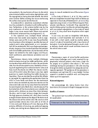
Percutaneous closure carries multiple challenges which include crossing the leaflet perforation, which can be done from either the LA side or the ventricular side, the site of transseptal access, feasibility of cross- ing and negotiating the mechanical aortic valve, and how much the device can affect the closure mecha- nism of the mitral valve. We chose a very low septal puncture to avoid stretching the leaflet during ma- nipulation. Then we decided to use either IM catheter or cut pigtail for negotiating the perforation from the LVOT as it was faster and easier. For the mechanical aortic valve, we avoided any excessive tension on the valve and made sure to stay away from the central slit to avoid impairment of both discs simultaneously. The best selection of the closure device was a double disc device with a distance no more than 4mm between the discs, and it is best to have a larger disc towards the high-pressure chamber (LV). Also, there must be enough distance between the edge of the device and the closure line of the mitral valve. We used an atrial septal occluder device size 4 mm with an LV disc of 12 mm and waist thickness of 3 mm. Because of the extreme difficulty of crossing, we preferred keeping a safety wire during device deployment to maintain
access in case of accidental loss of the access (Figure 3A-D).
In the study of Velasco S., et al. [3], they used an 8X4-mm Amplatzer Vascular Plug III with no follow up reported. In the study of Raczkiewicz S., et al. [4], they reported using a 6 mm × 3 mm PLD rectangular (Para- valvular Leak Device, Occlutech). They reported five months follow up by transthoracic echocardiography with no residual regurgitation. In the study of Javed U., et al. [5], they used 5mm Amplatzer atrial septal occluder.
In our case, we used an Amplatzer ASD device, however, a small Amplatzer duct occluder II, (5 to 6 mm with a short waist), could be another option since it’s made of micronitinol with a low chance of hemolysis. It can be delivered through a much small- er delivery sheath which could minimize trauma to the mitral valve as well.
Summary
Percutaneous repair of mitral leaflet perforation caries many challenges and is only reserved for ap- propriately selected patients who have a high risk for surgery or in patients who refuse it. The main challenge during the procedure is the safe crossing through the defect using both the antegrade and ret- rograde approach. TEE guidance of the procedure is mandatory and real-time 3D is very helpful. Further research is needed to establish mid- and long-term follow up of this approach.
Acknowledgment
We acknowledge Ms. Salma Elasfar from Cha- tham-kent, ON, Canada, for English language revision.
Conflict of Interest
The authors have no conflict of interest relevant to this publication.
Comment on this Article or Ask a Question
Journal of Structural Heart Disease, October 2018
Volume 4, Issue 5:234-239
