
Page 26 - Journal of Structural Heart Disease Volume 5, Issue 2
P. 26
39
Case Report
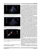
Video 1. The reversal of flow in descending aorta in diastole without aortic regurgitation. View supplemental video at https:// doi.org/10.12945/j.jshd.2019.014.18.vid.01.
Video 2. Shunt-like flow coming from aorta to pulmonary artery (transthoracic echocardiogram). View supplemental video at https://doi.org/10.12945/j.jshd.2019.014.18.vid.02.
Video 3. APF between the posterior right aspect of the aor- ta and the pulmonary artery (transesophageal echocardio- gram). View supplemental video at https://doi.org/10.12945/j. jshd.2019.014.18.vid.03.
sented to ER due to severe abdominal pain, mostly localized to right upper quadrant.
At the OSH ER abdominal CT was done which showed questionable injury to liver and spleen. At that moment it was decided to transfer the patient to our hospital for trauma evaluation. Following ar- rival to our hospital, abdominal CT was found to be reassuring. Upon further questioning it came to our knowledge that patient had multiple months of in- creased exercise intolerance including dyspnea on exertion which was occurring quicker into activities. Patient also reported orthopnea and was using an ex- tra pillow to sleep during this time. Given the afore mentioned, chest X-ray was obtained and found to be concerning for bilateral pleural effusions.
Cardiology was consulted due to positive history of complex congenital heart disease. Transthoracic echocardiogram (TTE) was completed and showed reversal of flow in descending aorta in diastole with- out evidence of aortic regurgitation (Video 1). Fur- ther investigation on echocardiogram made us sus- pect that there is shunt-like flow coming from aorta to pulmonary artery (Video 2), but it was not clearly identified on TTE. It was determined that a diagnostic catheterization and transesophageal echocardiogra- phy (TEE) would be needed to identify the source.
The patient had increased oxygen requirements due to worsening pleural effusions and increasing as- cites requiring transfer to the cardiac intensive care unit where bilateral thoracentesis was performed. Bilateral 8 Fr chest tubes were placed followed by al- most complete resolution of his bilateral pleural ef- fusions. However, prior to catheterization the patient had a persistent right sided pleural effusion causing desaturations while under anesthesia. Right chest tube (20 Fr) was placed which helped stabilize the pa- tient but failed to adequately drain the effusion. The patient was taken to the catheterization lab where surgical drainage was performed under general an- esthesia.
Once placed under anesthesia and the right sided pleural effusion drained, TEE was done. The TEE con- firmed our suspicion of APF between the posterior right aspect of the aorta and the pulmonary artery (Video 3). It was determined that a percutaneous ap- proach should be attempted with cardiothoracic sur- gery back up.
Right heart catheterization was performed using 7-Franch Berman angiographic catheter and revealed
Dzelebdzic S. et al.
Fontan Failure
