
Page 26 - Journal of Structural Heart Disease Volume 5, Issue 3
P. 26
65
Original Scientific Article
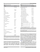
Table 1: Demographic Data Age
Sex
Male
Female
Diagnosis Tetralogy of Fallot Pulmonary stenosis PA/IVS
Pulmonary regurgitation Truncus Arteriousus Rheumatic heart disease Initial Surgery Transannular patch RV-PA conduit Pulmonary valvotomy Ross
Valve sparing repair Not known
None
Valve Placed
Yes
No
Reason for unsuccessful placement RVOT size
Coronary compression Improvement with angioplasty Valve Type
Melody
Sapien
Table 2: Baseline Measurement and Volumetric Data
Category
Count (%)
Mean (SD)
31.3 (9-56)
12 (52%) 11 (48%)
17 (74%) 2 (9%)
1 (4%)
1 (4%)
1 (4%) 1 (4%)
12 (52%) 5 (22%) 2 (9%)
1 (4%)
1 (4%) 1 (4%) 1 (4%)
17 (74%) 6 (26%)
4 (67%) 1 (17%) 1(17%)
14 (82%) 3 (18%)
RVOT Measurements (mm)
BWD in AP plane
BWD in lateral Plane
cSSFP coronal plane
cSSFP sagittal plane
MRA larger diameter
MRA smaller diameter 3D-SSFP MRI larger diameter 3D-SSFP MRI smaller diameter
MPA Measurements (mm)
cSSFP coronal plane
cSSFP sagittal plane
MRA larger diameter
MRA smaller diameter
3D-SSFP MRI larger diameter
3D-SSFP MRI smaller diameter
Right ventricular end-diastolic volume (ml/m2)
Right ventricular end-systolic volume (ml/m2)
Right ventricular ejection fraction (%) Pulmonary regurgitant fraction (%)
20.9 (4.5) 20.9 (5.1) 20.9 (4.1) 20.5 (4.7) 25.6 (4.5) 19.4 (5.0) 24.2 (4.1) 19.5 (3.9)
26.0 (4.0) 24.8 (6.) 29.5 (6.9) 24.0 (6.1) 29.6 (7.1) 23.3 (4.8) 118.8 (39.6)
66.4 (29.3)
47.8 (9.4) 32.3 (17.1)
Categorical variables expressed as count (%) Continuous variables expressed as mean (range). PA/IVS = Pulmonary atresia with intact ventricular septum; RV-PA = Right ventricle to pulmonary artery; RVOT = Right ventricular out- flow tract
Seventeen (74%) patients had a valve placed success- fully, four (17%) were not placed due to RVOT size. Fourteen (82%) of the 17 successfully placed valves were MelodyTM valves (Medtronic Inc, Minneapolis, MN), the other 3 (18%) were Sapien valves (Edwards
Variables are expressed as mean (SD). BWD = Balloon waist diameter; AP = Anterior-posterior fluoroscopy; cSSFP = stacked cine steady-state free pre- cession imaging; MRA = Magnetic resonance angiography; 3D-SSFP = whole heart self-navigated radial MRI
Life Science, Irvine, Ca). The average age at the time of the procedure was 31.4 years-old (9-56) and 12 (52%) were male. Tetralogy of Fallot was the most common lesion (n=17, 74%), the other diagnoses included pul- monary stenosis (n=2, 9%) and one each of truncus arteriosus, ventricular septal defect with pulmonary regurgitation, pulmonary atresia with intact ventricu- lar septum, and rheumatic heart disease (n=4, 16%). Table 1 contains full demographic data.
The average RVOT measurement was 20.9 ± 4.5 mm by BWD in the AP plane and 20.9 ± 5.1 mm in the lateral plane. The RVOT average measurement by cSS- FP was 20.9 ± 4.1 mm in the coronal plane and 20.5 ± 4.7 mm in the sagittal plane. The main pulmonary ar-
Kurtz J. D. et al.
MRI vs Balloon Waist for Pulmonary Valve
