
Page 24 - Journal of Structural Heart Disease Volume 5, Issue 3
P. 24
63 Original Scientific Article
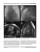
Figure 1. Examples of RVOT measurements. Panel A. BWD in AP fluoroscopy. Panel B. BWD in lateral flouroscopy. Panel C. cine steady- state free precession imaging (cSSFP) in oblique sagittal plane. Panel D. cSSFP in oblique coronal plane.
regurgitation [1]. The past two decades have seen the emergence of percutaneous valve replacement pro- cedures. This has given new options to those patients whom have undergone surgical and catheter based procedures on their PV and RVOT. Unfortunately with the current devices, the patient population eligible
for this procedure is limited by the size and shape of the outflow tract [2]. For the most part, these devic- es have been limited to right ventricle to pulmonary artery conduits or valve in valve replacements, with some use in native outflow tracts [3, 4].
Kurtz J. D. et al.
MRI vs Balloon Waist for Pulmonary Valve
