
Page 119 - Journal of Structural Heart Disease Volume 5, Issue 4
P. 119
181
Meeting Abstracts
patch with severe pulmonary regurgitation, right ventricle dilation and dysfunction, reduction in their NYHA class, cri- teria for pulmonary valve replacement.
Results: The implantation procedure was successful in all the patients resulting in an immediately functional valve. One patient had AV block that resolved after 24 hours. 1 valve had an infolding when implanted that resolved with balloon dilation, the valve mantained competente. Short term follow up, mean of 6 month (2-10) resulted in an improvement in NYHA class. Still no pulmonary valve regurgitation, reduction in right ventricle volumes from 150 ml/m2 (112-181) to 99 ml/m2 (78-119), less impres- sive reduction in ejection fraction on cardiac magnetic resonance. No stent fractures were observed at 6 months radioscopy.
Conclusion: Percutaneous pulmonary valve replace- ment with the Venus p-valve was safe and effective in our selected patients during the procedure and short time fol- low up.
155. TRANSCATHETER PERFORATION OF ATRETIC PULMONARY VALVE BY THE STIFF END OF A CORO- NARY WIRE IN NEONATES WITH PULMONARY ATRESIA WITH INTACT VENTRICULAR SEPTUM: A SOLUTION IN DEVELOPING COUNTRIES.
Sahar Elshedoudy, eman eldokla, Reem Rashed
Tanta University Hospital, Tanta, Egypt
Objectives: To evaluate the safety of using the stiff end of a coronary wire to perforate an atretic pulmonary valve (PV) in patients with pulmonary atresia with intact ventric- ular septum (PAIVS).
Background: Radiofrequency perforation is an accepted modality to perforate the PV in patients PAIVS. However, the high cost precludes its widespread use.
Patients and methods: This is a single-center experi- ence that spanned from March 2013 to January 2016 and involved 13 neonates who were severely cyanotic with PAIVS and with ductal-dependent pulmonary circulation.
The stiff end of a coronary wire was used to perforate the atretic PV anterogradely, followed by balloon pulmonary valvuloplasty.
Results: The mean age of patients was 3.9 ± 2.7 days and their mean weight was 2.8 ± 0.19 kg. The mean oxygen saturation was 77.1 ± 3.2%. All had membranous pulmo- nary atresia, with patent infundibulum and tripartite right
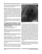
155. Figure 2. Controle angio by the end.
ventricle. The valve was successfully perforated in 11 out of 13 patients. Death occurred in two patients (15.4%) owing to heart failure and sepsis. Patent ductus arterio- sus stenting was performed 2 days after the procedure in one patient because of cyanosis followed by one and half ventricle repair at of age 5 months. Two patients (15.4%) had one and a half ventricle repair at age of 5 months and 6 months owing to insufficient anterograde pulmonary flow. Two patients (15.4%) underwent second intervention with balloon dilatation of the valve. The remaining seven patients (53.8%) had no further intervention. Two cases (15.4%) had femoral artery thrombosis treated with strep- tokinase. The mean duration of follow-up was 13.17 ± 7 months. There was significant improvement in the degree of tricuspid incompetence. There was a significant growth in the tricuspid valve annulus during the follow-up (the mean Z score increased from -0.8 ± 0.9 to 0.1 ± 0.9) (p = 0.003). There was also a significant increase in the tricus- pid valve annulus/mitral valve annulus ratio as its mean increased from 0.73 ± 0.10 to 0.86 ± 0.11 during follow-up (p < 0.001).
Conclusion: Perforation of the atretic PV in selected cases with membranous atresia and patent infundibulum using the stiff end of a coronary wire is an effective alternative to using radiofrequency perforation.
Hijazi, Z
22nd Annual PICS/AICS Meeting
