
Page 136 - Journal of Structural Heart Disease Volume 5, Issue 4
P. 136
Meeting Abstracts
198
airways. In several congenital lesions like the left pulmo- nary artery stenosis in hypoplastic left heart syndrome, information about vessel-airway relation may potentially impact treatment strategy. Outside the interventional car- diology space, tools have been developed to visualize (seg- ment) non-contrast regions in 3D rotational angiography (3DRA) such as biopsy or ablation targets.
Objectives: We investigated the feasibility of segmenting the airway from 3DRAs using commercially available tools.
Methods: A retrospective review of our institutional data- base was performed to identify all patients who underwent 3DRA with a new angiographic platform capable of airway segmentation (Philips Azurion, since 12/2018). Patients demographics and catheterization data were collected. Three-dimensional rotational angiographies including pul- monary arteries and airways were selected for further anal- ysis. A segmentation tool (XperGuide, Philips Healthcare) allowing for a stepwise 3D segmentation of structures with a low contrast ratio and irregular boundaries was used to present airway-vessel (pulmonary artery) relation. Three independent operators performed airway segmentation
from blinded 3DRAs. Segmentation time and completeness of segmented airway were recorded for all 3D datasets.
Results: Between 12/2018 and 4/2019, 3DRA was per- formed in 42 patients including 23 patients in whom pul- monary arteries were visualized. In the latter group the mean patient age and weight were 3.7 years (range: 1 month – 17 years) and 16.7 kg (3.9 – 85 kg), respectively. Seventeen patients presented with biventricular anatomy, the remaining 6 were single ventricle patients at various stages of palliation. Airway segmentation was performed successfully in 21 patients (21/23, 91.3%). In one patient the isocenter was set to anteriorly resulting in only par- tial inclusion of the airway in the 3D volume. In another patient artifacts from multiple previously implanted coils did not allow accurate visualization of the airways for seg- mentation. The median time for segmentation was 2:27 min (range 0:49 – 3:49 min) with no significant differences between the three operators (p=.9). Trachea was seg- mented in all patients (21), the left and right main bron- chus in 21 (100%) and 20 (95%) patients, respectively. Proximal right and left lobar bronchi were segmented in 17 (81%) and 11 (52%) patients, respectively.
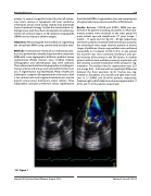
181. Figure 1.
Journal of Structural Heart Disease, August 2019
Volume 5, Issue 4:75-205
