
Page 138 - Journal of Structural Heart Disease Volume 5, Issue 4
P. 138
Meeting Abstracts
200
Introduction: The Ebstein anomaly covers less than 1% of cardiac malformations. The common feature of all cases is apical displacement and dysplasia of the septal valve of the tricuspid valve. Among the associated anomalies are the permeable foramen ovale or the atrial septal defect. 50% Interventricular communication or mitral valve disease.
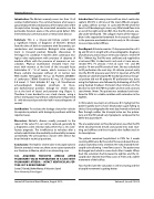
Summary: This is a 42-year-old female patient with a significant history of diagnosis of Ebstein's Disease from the time of birth in treatment with furosemide, spi- ronolactone and rivaroxaban. Biological valve replace- ment in tricuspid position Edwards Lifesciences 31 He began his condition 4 months after continuous bio- logical valve replacement with presence of dyspnea of medium efforts with the presence of murmurs on aus- cultation. Physical examination revealed heart mur- murs with murmur at the level of the tricuspid focus holisistolico IV / VI of Levine, which increases to the Rivero carballo maneuver without s3 s4 normal rest. With studies Hemoglobin 14.3 g / dl, Platelets 206,000 / ul Leukocytes 7.030ul Creatinine 0.5 mg / dl, Potassium 3.8 mmol / l Total bilirubin 0.7 mg / dl. Transthoracic echocardiogram biological prosthesis in tricus- pid dysfunctional position, leakage for severe valvu- lar at the level of lateral and posterior ring (Figure 1). Therefore it was decided to use a leak closure using a device during your stay without complications (Figures 2 & 3) with decreased paravalvular leak in ecocardiograma of control.
Justification: To evaluate the leakage closure for valvular tricuspidea in patients with biological prosthesis with few reported cases.
Discussion: Ebstein's disease usually proceeds to the repair of the valve if it can not be replaced, generally by a bioprotesic valve. Valvular replacement has a less satis- factory prognosis. The insufficiency to valvulate after a valvular substitution the morbidity and mortality increases considerably the percutaneous closure with device has been limited in technical questions.
Conclusions: The leak for severe valve in tricuspid valve in Ebstein anomaly is very rare, there are no cases reported in our literature in Mexico, which is an interesting case
182. ACQUIRED TRICUSPID ATRESIA AFTER PULMONARY VALVE PERFORATION IN A CASE WITH PULMONARY ATRESIA – INTACT VENTRICULAR SEP- TUM: IS IT A NEW FINDING?
Sonia El-Saiedi, Baher Matta, Al Hussein Sayed
Cairo University, Cairo, Egypt
Journal of Structural Heart Disease, August 2019
Introduction: Pulmonary atresia with an intact ventricular septum (PA IVS) is still one of the most difficult congeni- tal cardiac defects to treat. In cases with PA IVS when the heart is still developing in utero, very little blood flows into or out of the right ventricle (RV), thus the RV remains usu- ally under-developed. The collagen matrix of the myocar- dium is the supportive framework. An increase in collagen in the pressure-overloaded ventricle early in life is known to cause myocardial stiffness.
Case Report: A female neonate 17 days presented to us 3.5 kg and 50 cm in length with echocardiographic diagnosis of pulmonary atresia intact ventricular septum. Severe TR PG > 100mm Hg. The PAs were confluent and supplied by a tortuous PDA. Lt sided aortic arch and a 5 mm non-re- strictive PFO. TV annulus 7 mm (Z score -1.9) and MV annulus 14 mm and PA annulus 6 mm. She was scheduled to RF perforation the next day. JR catheter passed easily into the RV. RV pressure was125/4/14. Injection of the RV showed an underdeveloped RV mostly bipartite as the api- cal portion did not fill properly. RF perforation was done using the Nykanen wire followed by ballooning of the PV using Tayshak II balloon 7mmx2cm then 9mmx2 cm. The decision to stent the PDA was taken and we used coronary stent 4mmx 14mm. The patient was extubated and trans- ferred to CICU in a stable condition with saturation in the eighties.
In ICU patient was kept on primacore 0.5 mg/kg but the patient rapidly start to have desaturation again falling to sixties. Echocardiography the next day showed no forward flow through neither the tricuspid valve nor the pulmo- nary and the RV looked very hypoplastic compared to the ECHO the day before?!.
The one explanation to this phenomenon was that the RV when decompressed shrunk and passed into total stun- ning and stiffness and the tricuspid valve leaflets stuck in its closed phase.
The patient remained hospitalized in CICU for 2 weeks receiving Milrinone. By the end of the second week the sat- uration started to rise to seventies. Re-echo showed the tri- cuspid valve allowing 1 mm flow across it. The patient was discharged and asked to come back after 2 weeks. When back the saturation had increased to eighties and the tri- cuspid valve showed more flow across it 3 mm and a subtle movement of the leaflets.
Two months later the saturation is still increasing and the tricuspid movement is getting better.
Volume 5, Issue 4:75-205
