
Page 142 - Journal of Structural Heart Disease Volume 5, Issue 4
P. 142
Meeting Abstracts 204
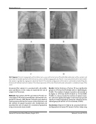
187. Figure 2. Panel A. Angiography of the inferior vena cava and Fontan tunnel. Panel B. After perforation of the conduit and atrial wall, the needle position within the atrial cavity is verified angiographically. Panel C. Exact placement of the stent within the septum is guided by angiograms obtained. Panel D. The desired configuration of the stent is achieved by slowly filling the balloon with diluted contrast medium. This allows expansion of the stent from both ends and results in a diabolo shape of the stent.
discovered that calpain-9 is associated with cell prolifer- ation and fibrosis. In this study, we examined the role of calpain-9 in PV stenosis.
Methods: Non-stenosis (Ns-PV) and stenosis PV with vas- cular stents (S-PV) were harvested from a patient with con- genital PV stenosis. H&E, Masson-Trichrome and calpain-9 staining were performed to measure intimal thickness and the number of calpain-9 positive cells. Furthermore, we examined the role of calpain-9 in neointimal hyperplasia using a mouse wire injury model.
Results: Intimal thickness of human PV was significantly greater in S-PV than Ns-PV (470.0±123.6 vs 128.8±40.2μm, P<0.01). The number of calpain-9 positive cells tended to be higher in S-PV than Ns-PV (16.6±6.2 vs 13.7±4.5/HPF, P=0.09). In a mouse model, the number of calpain-9 posi- tive cells were significantly higher in wire injure group with significant intimal hyperplasia formation compared to the control group (45.9±10.9 vs 16.7±5.9/intima, P<0.01).
Conclusions: Calpain-9 is likely to be associated with inti- mal hyperplasia in human PV stenosis. Further study using
Journal of Structural Heart Disease, August 2019
Volume 5, Issue 4:75-205
