
Page 89 - Journal of Structural Heart Disease Volume 5, Issue 4
P. 89
151 Meeting Abstracts
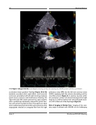
119. Figure 1 (Image 1A & 1B). Color Doppler showing scimitar vein draining into IVC/RA junction with flow acceleration.
acceleration (mean gradient=7mmHg) (Figure 1A & 1B), moderate secundum atrial septal defect, small muscular ventricular septal defect with left-right shunting, moderate patent ductus arteriosus with bidirectional shunting. The right ventricular (RV) systolic pressure was supra-systemic with a qualitatively moderately reduced RV systolic func- tion and normal LV systolic function. The patient was taken to the catheterization lab where the main pulmonary artery angiography revealed an antegrade flow from the right
pulmonary artery (RPA) into the left main coronary artery (LMCA) supplying both the anterior descending and the circumflex arteries (Figure 2). A selective injection within the anomalous scimitar vein showed drainage of the right lung into a curvilinear vertical vein connecting with steno- sis to IVC at the level of the diaphragm (Figure 3).
Role of Imaging in Patient Care: Imaging of the coro- nary origin in patients with ALCAPA can be challenging
Hijazi, Z
22nd Annual PICS/AICS Meeting
