
Page 90 - Journal of Structural Heart Disease Volume 5, Issue 4
P. 90
Meeting Abstracts
152
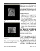
119. Figure 2. Injection into MPA showing LMCA origin from RPA.
119. Figure 3. Injection into a stenotic scimitar vein connecting into IVC/RA junction.
especially if the LMCA originates from RPA. Also, the pres- ence of pulmonary hypertension might contribute to maintain coronary perfusion and lead to misinterpretation of the antegrade flow in LMCA and its branches.
Cardiac catheterization remains the best imaging modal- ity to evaluate the coronary arteries preventing a poten- tially catastrophic outcome as demonstrated in this case. Catheter intervention with a series of balloon dilations of the stenotic scimitar vein was successful in relieving the stenosis. Moreover, coil embolization of two major collat- erals arising from the aorta and supplying the right lower posterior lung was performed. Then, the patient under- went reimplantation of LMCA into the aorta with a favor- able outcome.
Summary/Discussion Points:
• An extensive review of the available literature revealed only few cases of Scimitar syndrome associated with ALCAPA. In all of these cases, the LMCA originated from the posterior sinus of MPA. Our case is the first to report ALCAPA from RPA in association with Scimitar syndrome. This presentation might have led to the initial misinter- pretation of the echocardiography images.
• The presence of pulmonary hypertension in our patient maintained an adequate antegrade flow across the LMCA preventing significant coronary steal and signs of myo- cardial ischemia.
• The report highlights the essential role of cardiac cath- eterization not only assisting in the diagnosis and inter- vention on the scimitar vein but also to rule out poten- tial coronary arteries anomalies in patients with Scimitar syndrome, as this a rare although a very significant as- sociation that may have important implications in their outcomes.
120. COMPARISON OF PULMONARY ARTERY DIMEN- SIONS OBTAINED FROM CONVENTIONAL ANGI- OGRAPHY, 3D-ROTATIONAL ANGIOGRAPHY AND MULTI-SLICE COMPUTED TOMOGRAPHY
Ryan Pewowaruk , Alejandro ALZATE Roldan Alzate, Carolina Larrain, Christopher Francois, Luke Lamers
University of Wisconsin - Madison, Madison, USA
Introduction: Precise imaging of the pulmonary arteries (PA) is essential for management of patients with complex congenital heart disease (CHD). Conventional 2D angiog- raphy (CA) remains the gold standard for morphological and quantitative assessment of the PAs. Previous studies demonstrate strong correlations of measured PA diame- ters between CA and multi-slice computed tomography (MSCT). 3D rotational angiography (3DRA) has similar imaging capabilities to MSCT and has been used to guide PA interventions in CHD; yet no objective assessment of 3DRA image capabilities for PA anatomy exist and it is unclear how PA stenosis and PA stents influence 3DRA
Journal of Structural Heart Disease, August 2019
Volume 5, Issue 4:75-205
