
Page 29 - Journal of Structural Heart Disease Volume 5, Issue 5
P. 29
New Technology
222
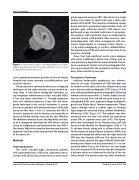
Figure 1. Cocoon device, which is an Amplatzer duct occluder I [ADO I] like device used for the closure of patent ductus occlud- er. [Reproduced with permission]
sure is preferred wherever possible in view of shorter hospital stay, easier recovery, cost-effectiveness, and cosmetic reasons.
The procedure is commonly done by an antegrade technique via the right ventricle, using an arterio-ve- nous loop. A trans-aortic retrograde technique us- ing Amplatzer membranous or duct occluder (ADO) II has also been described [1]. Though procedure time and radiation exposure is less with the retro- grade technique it has certain limitations. It cannot be used in patients with perimembranous VSDs with aortic rim less than 3 mm [1]. Secondly, the ADO II device is expensive and its routine use, especially in resource-limited settings may not be cost effective. We therefore aimed to assess the feasibility and safe- ty of a retrograde technique for VSD device closure using an indigenously manufactured low cost ADO I like device. We further aimed to demonstrate that this approach could be used irrespective of the adequacy of the aortic rim.
Methods
Patient population
This series included eight consecutive patients with perimembranous VSDs who underwent percu- taneous VSD device closure by the trans-aortic retro-
grade approach using an ADO I like device at a single tertiary care center in South India over a three year period (2015-2018). The hospital institutional review board and ethics committee approved the study. All patients gave informed consent. VSD closure was performed as per standard indications in symptom- atic patients, with significant shunt as evidenced by standard clinical (mid-diastolic flow murmur), elec- trocardiographic (left atrial enlargement, left ven- tricular hypertrophy) or imaging criteria (Qp/Qs > 1.5 by echocardiography or cardiac catheterization. Perimembranous VSDs with aortic rim less than 5 mm were also included.
Cases that had significant aortic valve prolapse with aortic insufficiency, defect size >10mm, and se- vere pulmonary hypertension were excluded. The pa- tients underwent clinical and echocardiographic fol- low-up one week after the procedure and then once every three months for one year.
Description of technique
Infective endocarditis prophylaxis was adminis- tered in all cases. Calculation of VSD diameter and its relation to the aortic valve was determined using trans-thoracic echocardiography (TTE) (Figure 2A & B) only without performing ventriculography. Following femoral arterial access with a 7 French sheath, the de- fect was crossed from the left ventricle using an ex- changeable 0.035-inch angulated floppy hydrophilic guide wire (Radio focus, Terumo cooperation, Tokyo, Japan) through either a Judkins right or Amplatz right diagnostic coronary artery catheter (Medtronic, Minnesota, USA) (Figure 3A). The catheter was then advanced over the wire into either the pulmonary artery (PA) or superior vena cava (SVC). The hydro- philic wire exchanged for an Amplatz extra-stiff soft tip wire (Cook Medical Inc. Bloomington, USA). A 7-8 Fr. delivery sheath along with its dilator which used for the closure of patent ductus arteriosus (PDA), was advanced through the defect into the right ventricle (RV) over the Amplatz stiff wire. The sheath was ad- vanced slowly towards the apex of the RV while with- drawing the dilator and was positioned 3–5 cm be- yond the defect (Figure 3B). A device 1 to 2 mm larger than the measured VSD diameter was pre-loaded on a delivery cable and advanced to the tip of the de- livery sheath. The entire system was then withdrawn
Journal of Structural Heart Disease, October 2019
Volume 5, Issue 5:221-228
