
Page 31 - Journal of Structural Heart Disease Volume 5, Issue 5
P. 31
New Technology 224
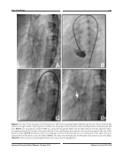
Figure 3. The steps of the procedure (an illustrated case): 0.035-inch angulated floppy hydrophilic guide wire, Terumo from the left ventricle through a Judkins right diagnostic catheter was advanced to the pulmonary artery exchanged with an Amplatz extra-stiff wire. Panel A. 7Fr. long delivery sheath of PDA was advanced through the defect into the right ventricle over the rigid wire which was advanced further to the apex of the right ventricle. It was confirmed by hand injection of contrast through the side arm of the delivery sheath. Panel B. Complete system was withdrawn with the exteriorized RV disc to be deployed on the right ventricular side and waist of the device across the defect. Panel C. The device was released after confirmation of its position across the defect (Panel D) by trans-thoracic ecocardiography which also rules out aortic regurgitation.
Journal of Structural Heart Disease, October 2019 Volume 5, Issue 5:221-228
