
Page 39 - Journal of Structural Heart Disease Volume 5, Issue 5
P. 39
Case Report
232
Figure 4. High coronary occlusion risk: Angiographically ob- served short VTC distance between left main ostium and surgical post. (A) Surgical frame post, (B) Left main artery, (C) Short left VTC distance.
The first strategy was selected. The plan was to use a 23 mm EDWARDS SAPIEN 3 balloon expandable valve instead of the 26 mm valve as suggested by the VIV application and CT analysis. Subsequently, this decision was selected due to the slightly longer VTC distance with a 23 mm valve to lower risk of coronary occlusion. Therefore, a 3.1 mm VTC distance was used versus the 2.6 mm. Doing this created less lateral dis- placement of the surgical posts with a smaller diame- ter THV valve. A shorter frame height valve of 23 mm was used instead of the 26 mm SAPIEN 3 valve. The sinotubular junction height in this patient is 16.4 mm, which is why the slightly shorter surgical frame post of 17 mm was selected. This would hypothetically cause less interaction of the surgical posts with the sinotubular junction having a shorter THV frame.
Slightly lower deployment was planned to mini- mize any potential interaction between the laterally displaced surgical posts and the sinotubular junction because of deployment of the THV valve. The left 2:1, right 2:1 and 1:1:1 fluoroscopic angles were identified and angiographic images were obtained. While using the XB 3.5 and JR4 guide catheters, coronary stents were placed in the mid LAD and mid RCA. There was a high likelihood a left main stent would need to be deployed to create a chimney path next to the THV stent frame. Aortic root angiography during a 23 mm balloon inflation in the left 2:1 angulation and the
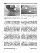
Figure 5. Coronary stent in a vertical trajectory against surgical aortic prothesis with the stent balloon can not be withdrawn back due to catheter entrapment. (A) THV. surgical prothesis frames. (B) Left main stent in a vertical trajectory against surgical bioprosthesis with deflated balloon entrapped inside the stent. (C) XB 3.5 guide catheter.
right 2:1 angulation confirmed the high coronary occlusion risk. Thrombolysis in myocardial infarction (TIMI) 3 flow was still maintained in both the left main and RCA during balloon inflation (Figure 4).
The 23 mm EDWARDS SAPIEN 3 valve was de- ployed in the left 2:1 view to evaluate the interaction between the left main ostium and the surgical valve prothesis during the THV valve deployment. Towards the end of the THV inflation period an interaction took place between the left coronary stent catheter shaft and the surgical posts/THV frame. Angiography post THV deployment showed the patient continued to have TIMI 3 flow in both the left and right coronary arteries. The wire and stent in the RCA were removed since there was no concern about RCA flow. However, due to the very short distance between the left main ostium and the surgical posts, deployment of the left coronary stent in and out of the left main coronary artery was performed. This created a vertical chim- ney appearance with a stented path extending to the level of the sinotubular junction. Easy future access could now be obtained if needed to the very low left main coronary artery, which had shallow sinuses and a short VTC distance of 3.1 mm.
Significant resistance was encountered while at- tempting to pull the stent catheter back from the parked mid LAD segment. While positioning the stent at the left main level, the stent catheter shaft was en-
Journal of Structural Heart Disease, October 2019
Volume 5, Issue 5:229-236
