
Page 41 - Journal of Structural Heart Disease Volume 5, Issue 5
P. 41
Case Report 234
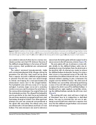
Figure 7. Panel A. Inability to wire through a superior strut due to distance and due to the presence of deflated winged balloon inside the stent. (A1) distal marker of stent balloon (A2) Coronary stent with deflated balloon entrapped inside. Panel B. Successfully wiring the stent through an inferior strut THV. (B1) wire #1 (B2) wire #2 introduced for better support (B3) distal marker of stent balloon.
was unable to advance further due to a narrow sino- tubular junction and short VTC distance. Presence of the deflated winged balloon in the lumen of the left main coronary stent prohibited wire advancement (Figure 7A).
The patient remained hemodynamically stable with no electrocardiogram (ECG) changes during the procedure. The left main stent could not be wired from a superior strut with a deflated winged balloon as planned. The left main stent was wired through an inferior strut hoping for a more permissible wire advancement course next to the winged balloon. The hydrophilic wire with two curves at its tip was reshaped. A primary larger curve and a secondary smaller curve was used to advance the wire freely in the narrow distance between the surgical frame and the sinotubular junction. This allowed the hydrophilic wire to go inferior to the stent frame and redirected the wire more superiorly to engage the left main stent from below, through an inferior strut. After significant attempts the wire was advanced and positioned in the apical LAD successfully. The inferior entry strut was dilated with a 2.0 mm balloon and then a 2.5 mm balloon. The same maneuver was used to advance a
second wire for better guide catheter support and to secure access to the left coronary arteries (Figure 7B). The only option left was to advance a snare cath- eter distally to the deflated balloon while also at- tempting to snare the balloon from the distal end in the proximal LAD. An attempt was made to retrieve the deflated balloon backwards, forcing it to make a short U-turn in the proximal lumen of the LAD. This would allow the dilated inferior left main strut to be removed without needing to pass in-between the surgical and the SAPIEN 3 frames. The distal end of the balloon was successfully removed by positioning a snare in the mid LAD and gradually pulling it back to capture the distal nose of the deflated balloon. A forceful pull was again applied, successfully retriev- ing the deflated balloon outside the left main stent
(Figure 8).
The existing left main stent will have a high risk
of thrombosis due to its malposition as described above. One way to minimize this risk was to reat- tempt to wire the left main stent from a superior strut now that the deflated winged balloon was removed and no longer in the way.
Journal of Structural Heart Disease, October 2019
Volume 5, Issue 5:229-236
