
Page 26 - Journal of Structural Heart Disease Volume 5, Issue 6
P. 26
Original Scientific Article
250
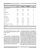
Table 1. Baseline clinical, echocardiographic and CT characteristics.
All (n=27)
No/trace PVL (n= 8)
Mild/gradeII PVL (n=19)
P value
Patients Characteristics
Age (years) Female BSA (mm2)
Echo Parameters
AVA (mm2)
Peak gradient (mmHg) LVEF (%)
CT Parameters
Mean annulus (mm) Annulus área (mm2) Annulus perimeter (mm) Mean LVOT (mmHg) Eccentricity index
Cover index
Moderate/severe valve calcification
Prosthesis diameter – CT mean annular diameter (mm)
85±4.3 22 (81%) 1.6±0.1
0.74±0.16 68±21 64±13
23±1.8 403±63 697±114 21.9±2.7 0.22±0.05 16.6±3.3 70% 4.6±0.9
83±2.4 7 (87%) 1.6±0.1
0.72±0.15 65±24 63±14
22.5±1.7 387±53 699±54 20.7±2.5 0.23±0.06 18.1±2.1 63% 5±0.5
86±4.8 15 (79%) 1.6±0.1
0.75±0.17 69±21 64±13
23.2±1.9 410±67 697±132 22.4±2.6 0.21±0.05 16±3.6 87% 4.4±0.9
0.1 0.6 0.5
0.7 0.6 0.9
0.4 0.4 0.9 0.1 0.5 0.07 0.4 0.04
Data are presented as mean ± standard deviation or as number (percentage) of patients. AVA = aortic valve area. BSA = body surface area. LVEF = left ventricle ejection fraction. LVOT = left ventricle outflow tract. NYHA = New York Heart Association; TAVI = Transcatheter aortic valve implantation; PPM = Permanent pacemaker
level of statistical significance was set at p ≤ 0.05. All statistical analyses were performed using SPSS Statistics version 18.
Results
Baseline clinical, CT and procedural characteris- tics are shown in Table 1. All 27 patients (mean age, 85±4 years, 81% of females) had severe aortic stenosis (mean AVA, 0.74±0.16 cm2). An iliofemoral access route was used in all the cases, and 33% of patients required balloon post-dilatation. Left bundle branch block was developed in five cases and a pacemaker was required in 15% of patients due to new conduction system ab- normalities. There were no deaths or major adverse cardiac events (MACE) during hospitalization.
Pre-discharge transthoracic echocardiography re- vealed mild or grade II PVL in 19 cases (70%) (16 pa- tients mild PVL; 3 grade II PVL). There were no patients with grade III or severe (grade IV) PVL. In all patients the regurgitation was paravalvular. Factors associated with the presence of PVL are presented in Table 1. The prosthesis/annulus discongruence (prosthesis diame- ter – CT mean annular diameter) was significantly re- lated to the occurrence of mild/grade II PVL (4.4±0.9 mm in the mild/moderate PVL group, versus 5±0.5 mm in the group without or trace PVL; p=0.04). However, no significant differences were found using area and perimeter values to calculate prosthesis/annulus dis- congruence. Moreover, no differences were observed when the body surface area (BSA) was taken into ac- count. Other factors such as eccentricity index, cover index, degree of annular calcification, mean annular
Journal of Structural Heart Disease, December 2019
Volume 5, Issue 6:248-253
