
Page 27 - Journal of Structural Heart Disease Volume 5, Issue 6
P. 27
251 Original Scientific Article
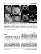
Figure 1. TOP: Panels A, B, C, D correspond to a patient without PVL after implantation of an Evolut PRO #29. Panel A. coronal CT image post TAVR shows mild oversizing with respect to the aortic annulus. Panel B. oblique transverse CT view show- ing a mean aortic annular diameter of 24 mm. Panel C. color Doppler in short axis view at the pre-discharge transthorac- ic echocardiography with no PVL. Panel D. Continuous wave Doppler through the aortic valve without any diastolic signal. BOTTOM: Panels E, F, G, H correspond to a patient with grade II PVL after implantation of a Evolut PRO #29. Panel E. coronal CT image post TAVR shows that TAVR is not oversized with respect to the aortic annulus. Panel F. oblique transverse CT view measuring a mean aortic annular diameter of 25 mm. Panel G. color Doppler in short axis view at the pre-discharge transthoracic echocardiography depicting a color jet around the aortic annulus of grade II PVL. Panel H. Continuous wave Doppler through the aortic valve with a continuous diastolic signal caused by the PVL.
and LVOT diameter, and baseline aortic regurgitation were not related to the occurrence of significant PVL.
Discussion
This study shows that PVL may still occur after TAVI using the novel CoreValveTM EvolutTM PRO sys- tem even when careful sizing, including systematic CT evaluation and technique, is used. In addition, our study demonstrates that the occurrence of mild or grade II PVL is related to a lower degree of oversiz- ing, measured as the disconcordance between pros- thesis diameter and mean CT aortic annular diameter (Figure 1). This novel insight may be clinically useful to optimize the implantation of this new device. In the Evolut PRO clinical registry, none to trace PVL was observed in 72.4% of patients while the remaining
27.6% experienced mild PVL at 30 days. There were no patients with moderate or severe PVL [5]. Com- pared with that study, the only currently available us- ing this valve, our study demonstrated the presence of mild or grade II PVL in 70% of the patients. The different criteria used for PLV assessment (systematic blinded echocardiographic review in our study) and patients characteristics (truly unselected cases in our study) could help to explain this apparently divergent findings.
The presence of significant PVL has been shown to be associated with worse short-term outcomes and increased in-hospital mortality [11]. In fact, the identi- fication of PVL predictors has been widely investigat- ed but with controversial results [12]. Consistent re- sults have been found by other authors with different valve systems. In patients treated with the Edwards
Alvarado Casas T. et al.
Paravalvular Leak Predictors Following TAVR
