
Page 28 - Journal of Structural Heart Disease Volume 1, Issue 3
P. 28
Original Scientific Article 134
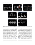
Figure 9: Pre deployment check: A. Leaflet insertion. B. Reduction in regurgitation (i) pre (ii) post. C. Pulmonary vein assessment (i) pre (ii) post. D. Mitral Stenosis (i) pre (ii) post.
echocardiographic views respectively (Figure 7). The trajectory of the clip is examined by moving the DC handle up and down while assessing the direction of the delivery shaft. Adjustments in the medial or lat- eral direction are made in the bi-commissural view either by moving the entire system or by adjusting the ‘M’ knob. Adjustments in the anterior or posterior direction are made in the LVOT view by rotating the guide handle clockwise or counter-clockwise. Once an ideal position is achieved, the clip is opened to 180 degrees and a 3-D en face surgical view of the mitral valve is obtained to assess for orientation of the clip arms relative to the leaflets (Figure 8). Any adjustments are made by rotating the DC handle in
the desired direction then transmitting the torque by moving the handle up and down rapidly. Once the clip is perpendicular to the leaflets, the clip is ad- vanced in the open position through the valve. The orientation of the clip is re-checked, the clip is closed to 120 degrees, and the DC handle retracted slowly to grasp both leaflets in the device. Once leaflet cap- ture is confirmed the grippers are pushed down and the clip is closed. TEE interrogation is then performed in multiple views to ensure leaflet capture with ad- equate tissue grasp, reduction in MR (assessed by regurgitant volume, size of MR PISA and pulmonary vein Doppler), and absence of a significant gradient (Figure 9). If these procedural goals have been met,
Journal of Structural Heart Disease, August 2015
Volume 1, Issue 3: 127-136
