
Page 26 - Journal of Structural Heart Disease Volume 1, Issue 3
P. 26
Original Scientific Article
132
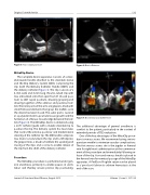
Figure 5: Trans septal puncture MitraClip Device
The complete device apparatus consists of a steer- able Guide handle attached to the steerable sleeve and the Clip Delivery System (CDS), comprising the clip itself, the Delivery Catheter Handle (DCH), and the delivery catheter (Figure 3). The clip consists of a 4-mm wide and 8-mm long chrome-cobalt clip with two articulated arms that open from 0° (closed posi- tion) to 240° (open position), allowing grasping and drawing together of the anterior and posterior leaf- lets. The inner parts of the arms are grippers, lined with small frictional elements that grasp the leaflets once the device has been closed. The outer part is covered in a polyester mesh to promote tissue growth and the formation of a fibrous tissue bridge between the leaf- lets (Figure 4). The MitraClip device is delivered using a 24 Fr catheter guide with a mobile steerable tip to position the clip. The delivery system has two knobs that control the anterior-posterior and medial-lateral steering of the catheter tip. The DC handle comprises two levers to lock/unlock the clip and to lift/depress the gripper lines, a knob to facilitate the opening and closing of the clips and a screw to enable release of the clip from the shaft of the delivery catheter.
Procedure
The MitraClip procedure is performed under gen- eral anesthesia, primarily to enable pauses in venti- lation and thereby ensure precise clip positioning.
Figure 6: Device Distance
Figure 7: Bicommisural and LVOT view
The additional advantage of general anesthesia is comfort to the patient, particularly in the context of extended periods of TEE evaluation.
One of the key advantages of the MitraClip proce- dure is venous access. We recommend using a micro- puncture needle to minimize vascular complications. The first venous access site is the jugular or femoral vein for right heart catheterization at the commence- ment of the procedure and immediately following re- lease of the clip. A second venous sheath is placed in the femoral vein for eventual passage of the MitraClip apparatus. A PerClose Proglide suture can be placed in a ‘pre-close’ fashion to achieve hemostasis at the end of the case.
Journal of Structural Heart Disease, August 2015
Volume 1, Issue 3: 127-136
