
Page 27 - Journal of Structural Heart Disease Volume 1, Issue 3
P. 27
133
Original Scientific Article
Cardiac imaging with visualization of the interatri- al septum (IAS) and the mitral valve apparatus is vital to the success of the MitraClip procedure. Operators should be well versed in obtaining and interpret- ing echocardiographic views to guide trans-septal puncture, device positioning and clip deployment. Furthermore, operators should be aware of the pa- rameters used to assess the success of clip deploy- ment based on echocardiographic interrogation. At our institution, TEE is performed by a cardiac anesthe- siologist experienced in MitraClip procedures, with an understanding of the expectations and requirements of the operator. Effective communication between the individual procuring the TEE images and the operator is imperative to facilitate an efficient and effective procedure.
The trans-septal puncture is arguably the most critical step of the procedure. If the puncture is inac- curate, subsequent device maneuverability and clip positioning is made difficult, often resulting in failed, or at best, unsatisfactory clip deployment position re- flected by minimal or no improvement in MR. Indeed, poor clip position may in fact worsen the degree of MR or cause MS. Accordingly, we take great care to ensure precise trans-septal puncture, repeating the process if necessary to ensure an optimal starting po- sition. The trans-septal puncture is performed under both fluoroscopic and TEE guidance using standard equipment and technique.
We recommend simultaneous viewing of a short axis image for anteroposterior positioning and a bi- caval image for superoinferior positioning. The opti- mal puncture site is located slightly inferior and pos- terior on the septum (Figure 5). Once this position is located we obtain a 0 degree, 4 chamber view to measure the “device distance”, defined as the dis- tance of the septal puncture from the mitral annu- lus. Ideally, this distance should be 4.0–4.5 cm above the mitral annulus as measured perpendicular to the plane of mitral valve coaptation during systole (Figure 6). If difficulty is encountered puncturing the septum, such as in the case of a thickened or fibrot- ic septum, focal cauterization of the septum can be used to facilitate entry.
Once the needle is across the septum the entire system is advanced into the left atrium and heparin is administered for anticoagulation. The 24F Abbott
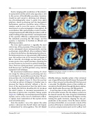
Figure 8: A. Checking orientation in 3D enface view. B. Correcting orientation in 3D enface view
MitraClip delivery steerable system is then advanced over a Superstiff wire into the left atrium. The Superstiff wire is then removed and baseline left atrial pressure is recorded. The MitraClip device is then carefully ad- vanced into the left atrium through the device deploy- ment sheath under fluoroscopy and TEE guidance.
From the plane of entry into the left atrium, paral- lel to the mitral annulus, the clip delivery system can be steered towards the valve using the mediolateral steering knob to turn the device 90 degrees and by turning the guide clockwise, aligning the clip per- pendicular to the annulus. Once the device reaches just above the leaflets, an assessment is made of the position of the clip in a mediolateral and anteroposte- rior plane, using bicommissural and LV outflow tract
Sharma, R.A. et al.
An Overview of the MitraClip Procedure
