
Page 19 - Journal of Structural Heart Disease Volume 3, Issue 1
P. 19
Original Scienti c Article 12
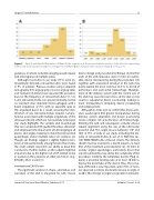
Figure 5. Panel A and Panel B. Distribution of (Panel A) the angulation of the proximal tubular portion of the left atrial appendage (LAA) to the horizontal plane (angle α, see Figure 3) and (Panel B) the change in LAA curvature (angle β, see Figure 3).
gulations, in whom complete imaging would require LAA interrogation in multiple views.
Although most LAAs in our study (77%) were an- terolaterally directed, retroverted LAAs were found in 9% of patients. Previous studies using computed tomography (CT) angiography, invasive angiography, and cadaveric materials have reported the presence, but not the frequency, of retroverted LAAs [15, 16]. Some retroverted LAAs can appear relatively normal on standard views (typically mid-esophageal, with a beam angulation of 75°), with an apparent apex at the angulated bend. As a result, ensuring the iden- ti cation of any retroverted lobes requires compre- hensive assessment with multiple angulations of the ultrasound beam, which are not routinely employed. Our study highlights the variable LAA morphology that can complicate TOE-guided thrombus detection and emphasizes the importance of LAA imaging in all planes and angles, keeping in mind that retroverted appendages and/or multiple lobes are common. Al- though our ndings suggest a slightly higher preva- lence of retroverted LAAs among females than males, the small sample sizes limit our ability to draw rm conclusions. Further studies on this subject might be important due to the higher risk of AF-related stroke in women in the presence of other risk factors (e.g., CHA2DS2-VASc score) [17].
Interventional LAA Closure
The signi cant variation in shape, orientation, and curvature of the LAA is important for LAA closure
device design and procedural technique. As the thin walls of the LAA (muscular wall ≤1 mm) are vulner- able, device maneuvering during the procedure [18] together with anatomical variation in the LAA may partly explain the most common (4–5%) [5, 6] risk of perforation and pericardial hemorrhage. Malalign- ment of the delivery system with the central axis of the LAA may cause tension/stress on the LAA or may result in suboptimal device positioning, leading to more manipulations including device recapturing and redeployment.
Although no trials exist to con rm this, many oper- ators would agree that greater manipulation during delivery system alignment and device positioning carries a higher risk of perforation and hemorrhage. Entering the LAA and subsequent occluder release require alignment along the axis of the LAA body/ proximal LAA. This angle (α) was between 110° and 140° in 75% of LAAs in our study, including the ma- jority of retroverted LAAs (6 out of 7). With the cur- rent WATCHMAN LAA occlusion device, the delivery sheath must be inserted to a depth equal to at least that of the maximum ostial diameter (21–33 mm). In acutely angled appendages, particularly those that are retroverted, this may add to the complexity of the procedure. Similarly, the Amplatzer Cardiac Plug re- quires a proximal landing zone of at least 10 mm to deploy the distal lobe of the device. Thus, the curva- ture of the LAA is also important, and in 58% of cases, we observed a mild to moderate increase in angle (1 to 60°). This change in angle was greater in retrovert-
Journal of Structural Heart Disease, February 2017
Volume 3, Issue 1:8-14
