
Page 18 - Journal of Structural Heart Disease Volume 3, Issue 1
P. 18
11
Original Scienti c Article
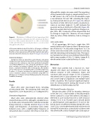
Figure 4.
although the sample sizes were small. The angulation of the proximal LAA (angle α) ranged widely, from 77 to 160° (mean, 125 ± 16°). In 75% of individuals, angle α was between 110 and 140°, including the majori- ty of retroverted LAAs (6 out of 7; Figure 5A). Almost two-thirds of LAAs (58%) had a mild or moderate in- crease in curvature (angle β, 1 to 60°) between the proximal and most distal part of the LAA (Figure 5B). Most retroverted LAAs had a signi cant change in an- gle (-30 to -90°). A minority of claw-shaped LAAs had no change in angle (8%). Likewise, all cone-shaped LAAs, by virtue of their de nition, had no change in angle.
Lobes and Lobules
Of all appendages, 66% had a single lobe, 30% were bi-lobed, and 4% were tri-lobed. The mean num- ber of lobes was 1.4 ± 0.6, with a range from 1 to 3. Six of the seven retroverted LAAs consisted of one lobe, with the remaining one having two lobes. The majori- ty of patients had at least one additional lobule (90%), with a mean number of 2.0 ± 1.2 lobules (range, 0–5), which tended to be located at the tip of a lobe.
Discussion
While most patients had a classical (i.e., claw- shaped) LAA shape and curvature, we found sig- ni cant variation in LAA orientation, shape, and curvature, with unusual shapes in 18% of patients, retroverted LAAs in approximately 10% of patients, and a wide range of angulations and multiple lobes in 90% of patients. This may have signi cant implica- tions for clinical practice during LAA closure or the identi cation of LAA thrombus by TOE.
TOE-Guided Thrombus Detection
Despite the utility of TOE for thrombus detection in the LAA [13], a small number of thromboembolic events continue to occur even when no thrombus is detected in a pre-cardioversion [8] or LAA-occlusion setting (1–2%) [5, 6]. Possible causes include absent or sub-therapeutic anticoagulation [14] and air em- bolism due to insu cient venting during LAA occlu- sion [5]. It is conceivable that thrombi may be missed during TOE examination, particularly in patients with retroverted LAAs and/or multiple lobes and large an-
Distribution of di erent left atrial appendage (LAA) positions/orientations. Anteverted (≤110°), anterolaterally direct- ed (111 to 150°), laterally directed (151 to 180°), or retroverted (>180°, with the major axis passing dorsally to a coronal section through the LAA ori ce).
of the main tubular body of the LAA or (2) lying in a di erent anatomic plane to the main tubular body. Lobules were de- ned by a clear separate pouch from the main lobe but with a division/cleft of the lobe by less than 50% (Figure 1B and 1D).
Statistical Analysis
Categorical data are reported as percentages, and group comparisons were performed using Fisher’s exact tests. Group comparisons for continuous data were performed using Wil- coxon-Mann-Whitney U tests. Normally distributed contin- uous variables (according to David’s test) are expressed as mean ± standard deviation (SD). In histograms, the number of class divisions was calculated by the root of the sample size. P-values < 0.05 were considered to indicate statistical signif- icance. Statistical analyses were performed using BiAS. for Windows 9.10 Software (Johann Wolfgang Goethe-University, Frankfurt, Germany).
Results
Shape
Most LAAs were claw-shaped (82%), including all retroverted LAAs. The remaining LAAs were fan- shaped (5%), cone-shaped (8%), or s-con gured (5%).
Orientation/Angulation
Most LAAs were anterolaterally directed when viewed from above (78%), although a sizeable minori- ty of LAAs were retroverted (9%), with the remainder being anteriorly or laterally directed (Figure 4). The ranges and frequencies of orientation were similar between sexes, with a slightly higher frequency of retroverted LAAs in women (16%) than in men (6%),
Joy, S. et al.
LAA Morphology in Non-Valvular AF
