
Page 17 - Journal of Structural Heart Disease Volume 3, Issue 1
P. 17
Original Scienti c Article
10
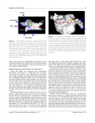
Figure 2. Three-dimensional surface-rendered image of the left atrium viewed from above (superior view) demonstrating the potential major directions of the left atrial appendage (LAA; blue arrows) relative to a coronal plane through the center of the left atrium (red dashed line). The LAA in this gure, marked by an asterisk (*), is in an anterolateral orientation. The green arrow indicates the angle of orientation measured from the coronal plane with zero in the right lateral position. LV, left ventricle; PVs, pulmonary veins; RUPV, right upper pulmonary vein; RLPV, right lower pulmonary vein.
tation, and lobes/lobules, rotating the image when necessary to obtain an optimal view. Further 2-D slices through relevant regions were used to determine other geometrical measures (e.g., angles) as detailed below.
LAA Morphology and Orientation Ascertainment
Shape. LAA shape was categorized into: claw-shaped, fan-shaped, cone-shaped, or s-con gured (Figure 1C). Cone- shaped LAAs narrowed to a tip at their distal point without any curvature, whereas claw-shaped LAAs had a single curve, and s-con gured LAAs had a double curve. Fan-shaped LAAs were thin and at, with the maximal width in one direction at least twice that in the perpendicular plane. The morphology of LAAs with complex shapes was categorized based on its closet resemblance to one of the four pre-de ned morphol- ogies. We preferred to use standard anatomical/geometric descriptions rather than pictographic descriptions (e.g., chick- en wing, cactus) due to inconsistencies in these pictographic shapes.
Orientation/Angulation. The orientation of LAAs was clas- si ed according to the major direction of the tip relative to the LAA ori ce in an axial plane. This orientation was de ned more precisely by determining the angle between a coronal plane through the center of the left atrium and the major lon- gitudinal axis of the LAA when viewed from above. Based on this angle, LAAs were assigned to the following categories: anteverted (≤110°), anterolaterally directed (111 to 150°), lat- erally directed (151 to 180°), or retroverted (>180°), with the major axis passing dorsally to a coronal section through the
Figure 3. Left atrium viewed from the anterior (ventral) surface demonstrating the angulation of the left atrial appendage (LAA), which is marked by an asterisk (*). The red dashed line indicates the horizontal (i.e., transverse) plane. The solid blue arrow indi- cates the direction of the proximal tubular portion of the LAA, and the orange blocked arrow indicates the angle between this and the horizontal plane (α). The blue dashed arrow indicates the line from the LAA origin to the tip, and the yellow blocked arrow indicates the change in angle between this and the solid blue arrow (β).
LAA ori ce (Figure 2). Multi-lobed LAAs (in which lobes often lie in di erent planes) were classi ed according to the orien- tation of the largest lobe. For twisted, multi-directional LAAs, analysis was performed in the direction of the major LAA part.
Precise measurement of LAA curvature is di cult; where- as identi cation of the long axis of the tubular neck is rela- tively easy, identi cation of the distal long axis is hindered by unclear tubular lines. We therefore examined the angulation of the proximal LAA neck (assessed as the angle between a transverse plane through the LAA ori ce and the major longi- tudinal axis of the proximal LAA (angle α, Figure 3) in addition to measuring the change in angle between this line through the proximal LAA and a line from the ori ce to the tip (angle β, Figure 3). These provide an indication of the degree of proxi- mal angulation and curvature of the rest of the LAA. In multi- lobed LAAs, analysis was performed on the major lobe. The change in angle was categorized as stable (0°), mild increase (1 to 30°), moderate increase (31 to 60°), or severe increase (>60°), with corresponding categories for negative values (su- perior/retroverted angulation).
Number of Lobes and Lobules. LAAs were considered to have at least one lobe (i.e., a tubular body with a blind-ending sac). If at least one cleft split the LAA by at least 50% of its length, the regions on either side of the cleft(s) were deemed to be separate lobes (Figure 1A). Further criteria, we took into consideration, were the ones de ned by Veinot et al. [9]: a vis- ible outpouching from the main tubular body of the LAA (usu- ally demarcated by an external crease) that was (1) occasion- ally but not necessarily associated with a change in direction
Journal of Structural Heart Disease, February 2017
Volume 3, Issue 1:8-14
