
Page 24 - Journal of Structural Heart Disease Volume 3, Issue 2
P. 24
Case Report
50
levels of triglycerides and cholesterol, consistent with a chylothorax. He was extubated shortly after tho- racostomy tube placement and was stable on room air, although a signi cant amount of chylous uid continued to drain from his thoracic cavity. A mag- netic resonance lymphangiogram was obtained to evaluate for congenital lymphatic malformation or signs of genetic lymphedema-lymphangiectasia syn- drome. However, imaging showed a normal thoracic duct with no obvious lymphatic malformation or in- trathoracic mass. An upper extremity vascular Dop- pler ultrasound was negative for deep venous throm- bosis in the right and left internal jugular, subclavian, and axillary veins. An echocardiogram had reportedly been notable for a patent foramen ovale and other- wise structurally normal heart at the referring hospi- tal and was repeated upon admission. Transthoracic echo showed a narrowed right-sided SVC with low velocity but forward ow to the right atrium. There was retrograde ow in the unobstructed innominate vein and multiple large posterior venous structures that appeared to be decompressing the upper body central veins caudally to a dilated inferior vena cava. The echocardiogram was negative for additional in- tracardiac or vascular lesions. The patient showed
no signs of plethora or upper body edema on exam. After the patient was stabilized, medical manage- ment was attempted, including octreotide infusion and low fat formula feeding followed by central line placement in the right subclavian vein and initiation of total parenteral nutrition (TPN) when the clinical response was deemed inadequate. Chylous drainage from the chest tube continued to be signi cant de- spite these measures. Given the echocardiographic nding of SVC obstruction as the likely etiology of continued chylous e usion, the patient was referred to the cardiac catheterization lab for angiography and intervention 14 days after admission.
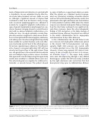
In the catheterization lab, the left femoral vein was accessed, and a hydrophilic catheter was placed in the stenotic SVC for hemodynamics and angi- ography. Right atrial pressure was normal, with a 5 mmHg gradient across the SVC. Angiography showed a severe long-segment SVC stenosis mea- suring 18 mm in length and 2.6 mm at its narrow- est diameter (Figure 1). The upper body central veins were decompressing through a very dilated hemiazygous system. The SVC was nearly occluded by the right subclavian vein central line that had been placed 3 days prior to catheterization. Balloon
Figure 1. Panel A. Severe long-segment superior vena cava (SVC) stenosis and collateral decompression of upper body venous ow through a very dilated hemiazygos system. Note that the SVC stenosis was appreciated prior to insertion of the right subclavian cen- tral venous line. Panel B. Lateral view, with thoracostomy tube in view.
Journal of Structural Heart Disease, April 2017 Volume 3, Issue 2:49-54
